Research in the Moody Lab
|
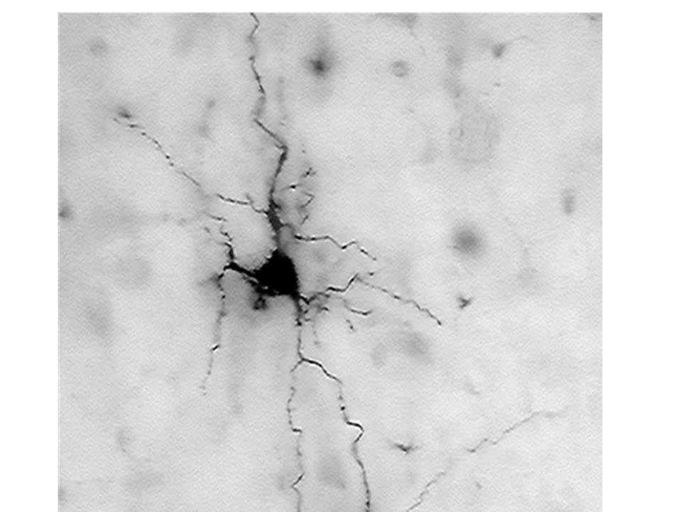
Above: Pyramidal neuron in a slice of neonatal mouse cortex, filled with biotin after voltage clamp experiment.
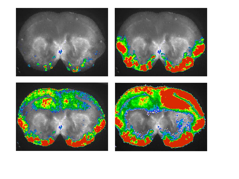
Above: Four frames from a film of a wave of spontaneous activity initaiting bilaterally in the ventral cortical pacemaker and propagating into the dorsal cortex. Fluo4-loaded coronal slice of P0 mouse cortex. Frames are at 0.5 sec intervals. |
Voltage- and ligand-gated ion channels show complex patterns of expression during the development of nerve and muscle cells. It is very difficult to reconcile this complexity with a simple linear approach of these cells to their mature physiological state. When viewed in light of the roles of spontaneous electrical activity in the development of nerve and muscle, however, the complex patterns of ion channel development make much more sense. Most nerve and muscle cells generate spontaneous electrical activity during at least one discrete stage of development and that activity plays fundamental roles in their development.
Our laboratory studies how the patterns of functional ion channel expression during development regulate spontaneous activity, and how that spontaneous activity in turn affects the later development of the cells.
There are currently two major projects in the laboratory:
1. A study of the development of voltage-gated ion channels in the embryonic and early postnatal mouse brain.
2. A study of highly synchronized, spontaneous electrical activity that occurs in mouse neocortex on the day of birth.
A third project, a study of ion channel development and spontaneous activity in ascidian (tunicate) muscle, formed much of the conceptual framework for our current mouse brain work. The ascidian muscle project is in hiatus for the present, but is likely to be restarted at a later date.
For a review of the field of ion channel development and spontaneous activity, you can read:
Moody WJ & Bosma MM. Ion channel development, spontaneous activity, and activity-dependent development in nerve and muscle cells. Physiol. Rev. 85, 883-941 (2005).
For recent work from our group, you can read:
McCabe, A.K.,Chisholm, S.L., Picken-Bahrey HL, & Moody, W.J. (2006). The self-regulating nature of spontaneous synchronized activity in developing mouse cortical neurones. J. Physiol. 577:155-167.
McCabe, AK,Easton, CR, Lischalk, J, & Moody, WJ (2007). Roles of glutamate and GABA receptors in setting the developmental timing of spontaneous synchronized activity in the developing mouse cortex. Dev. Neurobiol. 67:1574-1588.
Lischalk JW, Easton CR, Moody WJ. (2009). Bilaterally propagating waves of spontaneous activity arising from discrete pacemakers in the neonatal mouse cerebral cortex. Dev. Neurobiol. 69, 407-14.
Currie DA,Corlew R, deVente J, Moody WJ. (2009). Elevated glutamate and NMDA disrupt production of the second messenger cyclic GMP in the early postnatal mouse cortex. Dev. Neurobiol. 69:255-266.
Conhaim J,Cedarbaum ER,Barahimi M,Moore JG,Becker MI, Gleiss H, Kohl C, Moody WJ. (2010). Bimodal septal and cortical triggering and complex propagation patterns of spontaneous waves of activity in the developing mouse cerebral cortex. Dev. Neuro. 70:679-692.
Last modified:Oct. 2010