Moody Laboratory: Ion channel development in mouse cortical neurons
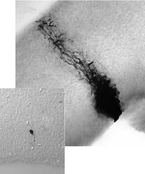
Many of the neurons of the neocortex are generated during embryonic development from a stem cell population in the ventricular zone (VZ). We studied the electrical properties of these VZ cells in mouse using whole-cell voltage clamp methods. Injection of the dye Neurobiotin, which crosses gap junctions, typically stains a large coupled cluster of VZ cells, including those of radial glial morphology (see Figure at left). Blocking gap junctions with octanol blocks this dye coupling. Experiments in rat VZ have shown that this extensive dye coupling is accompanied by strong electrical coupling. Our experiments in mouse show that although dye coupling in mouse VZ is similar to that in rat, electrical coupling in mouse is quite weak. This allowed us to voltage clamp mouse VZ cells accurately, without resorting to uncoupling methods.
These voltage clamp experiments showed that virtually all VZ cells express large delayed K currents. A small fraction of VZ cells also expressed inward Na currents. The change in this fraction during development suggests that VZ cells that express Na currents are those that have just exited the cell cycle and are in the process of migrating outward toward the cortical plate. Na currents are expressed in VZ cells of radial glial morphology as well as those that do not extend processes out of the VZ. As VZ cells migrated into the intermediate zone and cortical plate, the Na currents become larger but the outward K currents do not change.
You can read about this work in:
Picken-Bahrey, HP and Moody, WJ. Cerebral Cortex 13, 239-251 (2003)
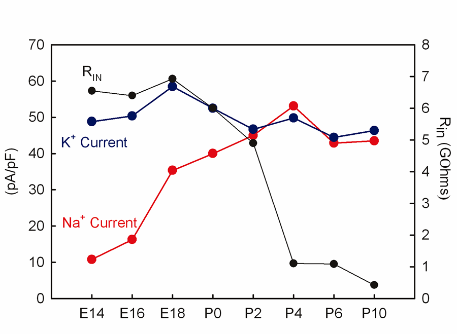
We have also studied the development of Na and K currents in cortical plate neurons through the embryonic and early postnatal periods. As shown the graph to the left, Na current density in cortical plate neurons increases about 4-fold between E14 and P2. Delayed K current density, in contrast, does not change during this period. This means that when a VZ stem cell exits the cell cycle, it beging to express functional Na currents, which then increase steady throughout early development. Delayed K currents, on the other hand, are present in VZ stem cells and do not change in density after cell cycle exit, or during migration and early differentiation in the cortical plate. The increasing ratio of inward Na to outward K currents would be exprected to increase neuronal excitability during this time. This is confirmed both by current clamp recordings showing marked increases in excitability and repetitive firing ability between E14 and P10.
The resting input resistance of embryonic cortical plate neurons is very high, averaging more than 5 GOhms. Immediately after birth (P0), input resistance falls dramatically, by more than 10-fold in the first 10 postnatal days. This would be expected to lower excitability and reduce synaptic strength.
The window of development between about E16, when Na currents begin to increase, and P4, when input resistance decreases, is a likely period of spontaneous activity in cortical neurons. We have confirmed the existence of spontaneous, highly synchronized activity at these stages in Ca imaging experiments.
You can read about this work in:
Picken-Bahrey, HP and Moody, WJ. J. Neurophysiol. 89, 1761-1773 (2003).
Last modified:Oct. 2010