Moody Laboratory: Spontaneous Waves of Activity in the Neonatal Mouse Brain
Many structures of the central nervous system generate spontaneous activity during early development and it is known that this activity is essential for a large number of important developmental processes such as neuronal migration, synaptogenesis, and the correct maturation of ion channel and receptor properties. We have discovered that spontaneous activity in the neonatal cerebral cortex occurs as propagating waves, which can be measured in wide-field high-speed calcium imaging in living brain slices. Films of this activity reveal that it originates in one of two bilateral pacemaker structures: a region of the ventrolateral cortex ventral to the rhinal fissue, or the septal nuclei. Surgical excision of these structures prevents activity. We are currently working to identify the pacemaker neurons and determine what properties allow them to initiate waves of activity.
You can read about this work in:
Lischalk JW, Easton CR, Moody WJ. (2009). Bilaterally propagating waves of spontaneous activity arising from discrete pacemakers in the neonatal mouse cerebral cortex. Dev. Neurobiol. 69, 407-14.
Conhaim J,Cedarbaum ER,Barahimi M, Moore JG, Becker MI, Gleiss H, Kohl C, Moody WJ. (2010). Bimodal septal and cortical triggering and complex propagation patterns of spontaneous waves of activity in the developing mouse cerebral cortex. Dev. Neuro. 70:679-692.
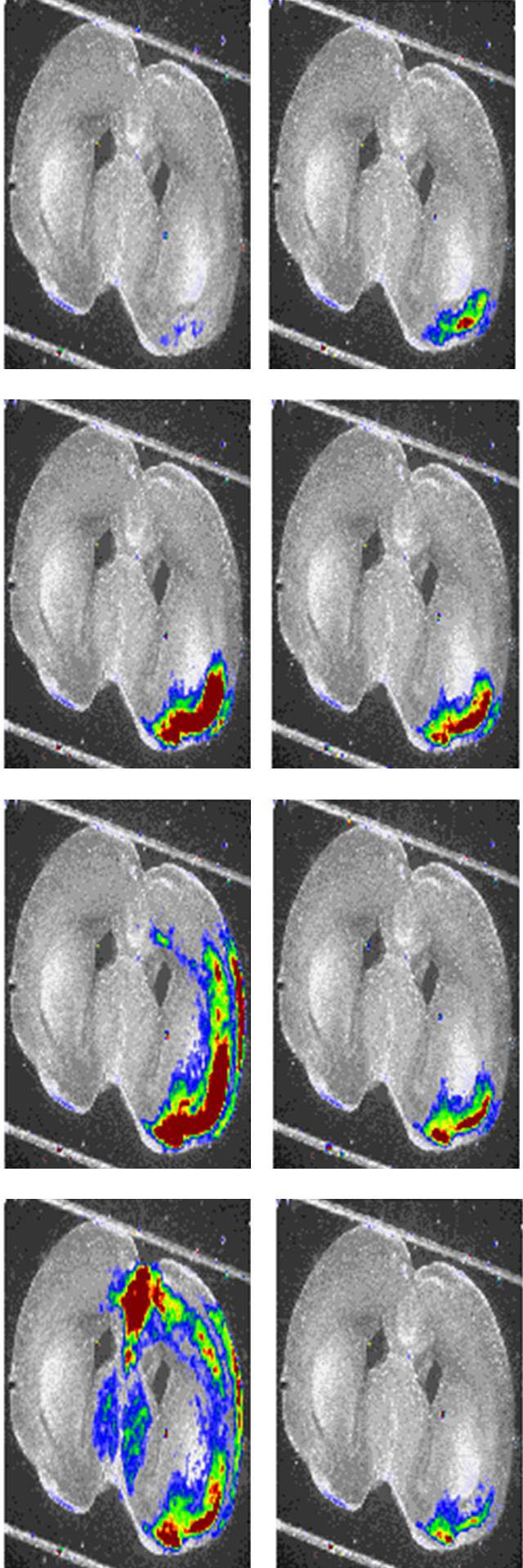
We have found the spatial patterns of wave propagation change dramatically with early development. At E18 (embryonic day 18; 1 day before birth), more than 90% of all waves are restricted to the ventral pacemaker region and septal nucleus. These waves stop at a very stereotyped point at the boundary between ventral and dorsal cortex. Just a few days later, at P5, the majority of waves propagate into the dorsal cortex. These waves slow transiently at exactly the same point that E18-P0 waves stopped - at the border between ventral and dorsal cortex.
Pharmacological evidence indicates that at earlier stages, waves are initiated by GABAergic neurons whereas at later stages they are more glutamate-dependent.
Last modified:Oct. 2010