Moody Lab: Ascidian muscle research
|
Figure 1: Embryos of the ascidian Boltenia villosa at five different stages of development. The endogenous orange pigment marks the muscle lineage cells. Below: Voltage-clamp records of the inward rectifier from muscle-ineage cells at each stage, showing its transient disappearance just after gastulation. |
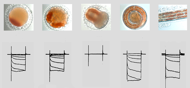
Ascidians are marine invertebrate chordates that have been a classic preparation in the study of development for many years. Their embryos have relatively few cells, develop rapidly, and many cell types develop without interactions with neighboring cells. The embryo of a local Puget Sound species, Boltenia villosa, has muscle-lineage cells that have an endogenous orange pigment, so that they can be identified at all stages of development, even before they express overt characteristics of muscle. Five stages of Boltenia embryos are shown in the figure: fertilized egg, gastrula, tailbud, pre-hatch tadpole, and post-hatch tadpole (only a portion of the tail is shown). These photographs span 40 hrs of development.
This pigment allowed us to identify, record from, and voltage clamp muscle-lineage cells at all stages of their development. We discovered that these cells generate spontaneous action potentials for a period of 6-8 hrs just after gastrulation, even when they are completely isolated from the embryo.
What triggers this activity? The voltage clamp records in the figure above show the inwardly rectifying K current, which is the only resting conductance in these cells, disappears for 6-8 hrs starting just after gastrulation. This destabilizes the resting potential, causing the cells to generate spontaneous bursts of action potentials.
What is the function of this spontaneous activity? Normally in these cells, the action potential shortens in duration by about 8-fold as the cell matures. This shortening must occur if mature muscle is to contract and relax at the high frequencies required for larval swimming. The shortening occurs because of the expession late in development of a rapidly activation Ca-dependent K current. If spontaneous activity is prevented, the Ca-dependent K current is not expressed and the action potential never shortens.
You can read about this work in the following papers:
Greaves AA, Davis AK, Dallman JE, and Moody WJ. Development of ionic currents in the muscle lineage of the ascidian Boltenia villosa. J. Physiol. 497:39-52, 1996.
Dallman JE, Davis AK, and Moody WJ. Spontaneous activity regulates calcium-dependent K+ current expression in developing ascidian muscle. J. Physiol. 511:683-693, 1998.
Dallman JE, Dorman J, and Moody WJ. Action potential waveform voltage clamp reveals the significance of the patterns of ion channel development in ascidian muscle. J. Physiol. 524:375-386, 2000.
Last modified: 4/05/2005 6:09 PM