| Conjoined Twins Separated |
| Conjoined Twins Separated |
|
By Ellen Kuwana Neuroscience for Kids Staff Writer August 14, 2002 Updated: October 13, 2003 On Monday, August 5, 2002, Guatemalan twins who were born joined at the head underwent more than 22 hours of surgery at Mattel Children's Hospital at the University of California Los Angeles (UCLA) to be separated. The girls were joined at the top of the head such that they faced opposite directions and consequently could not sit up. The twins, Maria de Jesus and Maria Teresa, just turned a year old. The UCLA doctors describe them as "amazingly pleasant" and "sweet, adorable." Maria Teresa is the stronger of the two and is starting to want to crawl. The girls were brought to the United States in May by an international charity named "Healing the Children." In mid-June, surgeons inserted eight-inch silicone balloons under the skin of their scalp in order to expand the skin. This process, which usually takes two to three weeks, was necessary to produce enough extra skin to cover the wound after separation surgery. It is important for the wound to be covered by skin to reduce the risk of infection. | 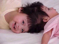 Photo courtesy of Mattel Children's Hospital, UCLA. Copyright 2002, University of California, Los Angeles |
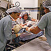 Photo courtesy of Mattel Children's Hospital, UCLA. Copyright 2002, University of California, Los Angeles |
Everyone Prepares for the SurgeryOnce the skin was expanded, the twins were ready for the surgery. How did the surgeons prepare for the complicated surgery? Using imaging technologies such as MRI and CT, the surgeons found that the twins' brains were separated by a membrane and seemed to function independently. That was good news because it meant that the twins did not share much brain tissue, which would make the surgery more straightforward. Each twin's brain appeared to be normal in size and structure. Each had normal arteries, the vessels that bring oxygenated blood to the brain. The surgery's main obstacle would be dealing with the sagittal sinus, a large vein that runs over the top of the skull from front to back. This major vessel carries blood from the brain back to the heart, where it is reoxygenated. The twins shared this important vessel. The surgeons predicted that they might have to give the vessel to one of the twins and hope that the other twin would be able to compensate using smaller vessels. This would put one twin at risk for a stroke. |
Surgeons Practice with Brain ModelsAs with any intricate procedure, it's best if one can practice first. Replicas of the twins' skulls were made using the information from the imaging studies. These three-dimensional, life-size plastic models helped the surgeons to familiarize themselves with the twins' unique anatomy: veins and arteries, shape of skull, relative placement of structures, etc. From the model, surgeons noted that the twins' heads and face were flattened on the left side and their ears were not aligned. The model could be viewed from various angles, different approaches mapped out and tested, alternative routes discussed and considered.
The SurgeryOn the day of surgery, the twins were positioned so that one was facing up and one was facing down, her head cradled in a donut-shaped pillow. At 8:00 a.m., the girls were wheeled into the operating room after getting kisses from their parents. The delicate procedures of preparing for surgery and getting the girls anesthetized began. The actual surgery started around 2 p.m. Bones and blood vessels had to be teased apart, the tissue separated, and then the equally delicate task of putting each girl back together began. Fortunately, the sagittal sinus was not as big of a problem as anticipated. The surgeons were able to check each girl's blood flow in various vessels to insure that each girl had sufficient blood supply. A new sagittal sinus did not have to be created, because the smaller vessels were able to compensate adequately.After almost an entire day (and night) of surgery, the girls were separated around 1 a.m., and at 5:45 a.m. the surgery ended. Still sedated, but free to move independently of each other for the first time in their young lives, the girls were monitored in the pediatric intensive care unit. |
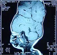 Photo courtesy of Mattel Children's Hospital, UCLA. Copyright 2002, University of California, Los Angeles |
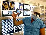 Photo courtesy of Mattel Children's Hospital, UCLA. Copyright 2002, University of California, Los Angeles |
A ComplicationFour hours after the surgery, Maria Teresa developed a build up of blood on the surface of her brain, and had to be brought back to surgery to relieve the pressure caused by the pooling blood. This complication is called a hematoma and is caused by bleeding from the veins around the brain. Maria Teresa had a subdural hematoma, meaning the blood was under the dura. The dura is a membrane that covers and protects the brain. The pressure from a large hematoma can damage the brain, so it has to be drained away. |
A Team EffortNeurosurgeons at UCLA had never separated conjoined twins before. More than 50 medical personnel volunteered their time and expertise. Among them were four pediatric anesthesiologists, two teams of plastic surgeons, and two teams of neurosurgeons, led by Jorge Lazareff, MD, and Henry Kawamoto, MD, DDS, who specialize in craniofacial surgery. UCLA has received around $4000 in donations from the public; costs are estimated at more than $1.5 million for the twins' medical care.Conjoined twins are rare. Conjoined twins joined at the head are the rarest of all -- only 2% of conjoined twins are joined at the head. Worldwide, only six twins joined at the head have been separated in the last 10 years. The most recent was in Singapore in April of 2001. In most cases, the separated twins have some deficits after the surgery and many require special education and rehabilitation. Another set of (Egyptian) twins who are joined at the head wait for their separation surgery, which will take place in Dallas, Texas; the surgery date is still unscheduled.
January 14, 2003The Maria twins are finally home -- after seven months in the hospital. They were scheduled to leave the hospital in late December 2002, but Guatemalan medical personnel needed additional time to get ready for the twins' arrival and ensuing health care needs. The girls' recovery is proceeding smoothly. They will continue physcial and occupational therapy in Guatemala. The girls are now 17 months old. |
Surgery is complete!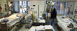 Photo courtesy of Mattel Children's Hospital, UCLA. Copyright 2002, University of California, Los Angeles |
September 2, 2003Maria Teresa has a hearing aid to help with hearing loss that was discovered after the August 5, 2002 surgery. The twins are still living in Guatemala, where UCLA surgeons visit from time to time. In April of 2003, surgeons visited the twins to deal with an infection that Maria Teresa was fighting. She had E. coli meningitis that was treated with antibiotics. (More about meningitis from the Centers for Disease Control and Prevention.) A month later, the girls returned to UCLA's Mattel Children's Hospital for an evaluation. While at UCLA, Maria Teresa, as a result of the meningitis, had to have a replacement shunt placed in her brain to drain excess fluid away from her brain. The Mattel Children's Hospital explains:
"As a result of last year's separation surgery, the cerebral spinal fluid in Maria Teresa's brain cannot flow normally through the drainage veins at the base of her head. UCLA doctors installed the shunt to help her veins manage the extra fluid. When Maria Teresa contracted E. Coli meningitis in Guatemala in April, the shunt became infected." On July 25, 2003, at UCLA, the girls celebrated their second birthday. Now both girls are in stable health and continuing to develop, although Maria Teresa's development trails that of her sister. Both continue physical therapy and occupational therapy. In the future, both will need more plastic surgery to "improve the shape of their skull caps and normalize their hair patterns," according to lead plastic surgeon Terry Kawamoto of UCLA. |
Twins leave the hospital. Photo courtesy of Mattel Children's Hospital, UCLA. Copyright 2003, University of California, Los Angeles |
Iranian Conjoined Twins Laleh and Ladan Bijani Die During Separation SurgeryUnfortunately, not all separation surgeries end successfully. On July 8, 2003, the 29-year-old Bijani sisters died from uncontrollable bleeding after the surgery. This surgery marked the first time surgeons have attempted to separate adult conjoined twins. In this case, the surgery was made more difficult because Laleh and Ladan were joined at the head. The sisters, who were law school graduates, wanted to be separated and had sought out doctors around the world. A medical team in Singapore agreed to perform the surgery. The women were aware of the risks involved, and felt that the complicated surgery would be worth undergoing in order to exist as two separate people. |
Egyptian Conjoined Twins Separated in Dallas, TexasMohamed and Ahmed Ibrahim, born June 2, 2001, in Egypt, arrived in Texas on June 22, 2002. In April of 2003, the boys had tissue expander surgery to create enough skin to accommodate a separation surgery. Separation surgery was completed on October 12, 2003. | 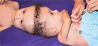 Mohamed and Ahmed Photo credit: World Craniofacial Foundation |
| Did You Know? |
|
|
References:
|
| GO TO: | Neuroscience In The News | Explore the Nervous System | Table of Contents |
![[email]](./gif/menue.gif) Send E-mail |
 Fill out survey |
 Get Newsletter |
 Search Pages |
 Take Notes |