| Twins Born Joined at their Heads are Surgically Separated |
| Twins Born Joined at their Heads are Surgically Separated |
| By Ellen Kuwana Neuroscience for Kids Staff Writer April 24, 2001
Twins Born Joined at their Heads are Surgically Separated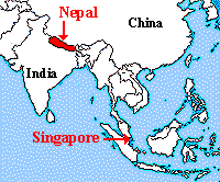 After 88 hours in the operating room, 11-month-olds Jamuna and Ganga are
two separate babies. The conjoined twins (also known as "Siamese twins")
from a mountain town in Nepal were sent to Singapore for surgery.
Conjoined twins are always identical and usually must be separated in
order for them to survive.
After 88 hours in the operating room, 11-month-olds Jamuna and Ganga are
two separate babies. The conjoined twins (also known as "Siamese twins")
from a mountain town in Nepal were sent to Singapore for surgery.
Conjoined twins are always identical and usually must be separated in
order for them to survive.The twins were joined at the tops of their heads, so that they were facing away from each other. They shared a brain cavity and their brains had fused partially. The hundreds of intermingled blood vessels in their brains made the surgery especially challenging. Neurosurgeon Keith Goh characterized the intertwined vessels as "torturous routes up a mountain." Another challenge was identifying and trying to preserve the speech and logic centers in each girl's brain. Although it was impossible to avoid some damage to the brains, surgeons did not want to injure these crucial areas. At this point, doctors do not know if either girl will have functional problems as a result of the surgery.
It is too soon to tell if the girls have any neurological damage. The biggest risk right now to the girls' recovery is infection and hydrocephalus, a condition in which the production and/or drainage of cerebrospinal fluid (CSF) is disrupted and too much fluid accumulates in the brain. The girls will probably stay in the hospital for the next three months. As of Monday, April 16, 2001, when doctors took off the girls' bandages, each girl had a fever and a high white blood cell count, both signs of infection. Doctors are pleased with the girls' progress, however, and optimistic about their recovery.
Conjoined twins are rare, but ones joined at the head are extremely rare, occurring approximately once every two million births. The first successful separation (meaning no neurological damage) of twins joined at head was performed in 1987. In the past 500 years, only about 80 cases of conjoined twins joined at the head have been known worldwide. Back home in Nepal, relatives gathered around their neighborhood's only phone, waiting for news. Having been told that the babies had a 50-50 chance of surviving the surgery, the family is calling the promising outcome a "miracle." As medical technology advances, perhaps more miracles will be possible. References:
|
| BACK TO: | Neuroscience In The News | Table of Contents |
![[email]](./gif/menue.gif) Send E-mail | ![[survey]](./gif/menusur.gif) Fill out survey | ![[newsletter]](./gif/menunew.gif) Get Newsletter | ![[search]](./gif/menusea.gif) Search Pages |
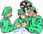 A team of 20 doctors took shifts, operating for almost four days. At any
one time, there were up to 16 doctors in the room. The surgeons had
expected the surgery to take about 40 hours but the surgery took much
longer because every blood vessel had to be traced and separated. One
consulting surgeon described the blood supply as looking like a "tangled
serving of spaghetti." The surgery was finished by plastic surgeons who
closed the skulls. Gortex, a synthetic material used in raincoats, was
substituted for the
A team of 20 doctors took shifts, operating for almost four days. At any
one time, there were up to 16 doctors in the room. The surgeons had
expected the surgery to take about 40 hours but the surgery took much
longer because every blood vessel had to be traced and separated. One
consulting surgeon described the blood supply as looking like a "tangled
serving of spaghetti." The surgery was finished by plastic surgeons who
closed the skulls. Gortex, a synthetic material used in raincoats, was
substituted for the 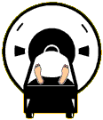 The doctors prepared for six months before the actual surgery. They
practiced "virtual surgery" with a three-dimensional imaging system using
pictures of the girls' skull cavities. The "3-D" images were made using
already existing
The doctors prepared for six months before the actual surgery. They
practiced "virtual surgery" with a three-dimensional imaging system using
pictures of the girls' skull cavities. The "3-D" images were made using
already existing