Neuroscience For Kids
Visual Pathway
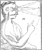 (Image courtesy National Library of Medicine, Bethesda, Maryland.) |
If you can understand the lower figure on this page, then you have learned the major pathway that visual information takes on its way from the eye to the primary visual cortex. The figure is really not too difficult to understand....really. Imagine that the colored bar (half red, half blue) is in front of your eyes. The red part of the bar will project to the nasal part of your left retina and the temporal (lateral) part of your right retina. The blue part of the bar will project to the nasal part of your right retina and the temportal (lateral) part of your left retina.
Follow the blue and red lines from the eyes to see the flow of information. From the retina, the first synapse is in the lateral geniculate nucleus of the thalamus. The next synapse is made in primary visual cortex in the occiptal lobe. |
 Right and left visual
information cross to opposite sides of the brain. This crossover occurs
in the optic chiasm. After the optic chiasm, information about the right
visual field (blue) is on the left side of the brain, and information
about the left visual field (red) is on the right side. The pathways stay
this way all the way up to the visual cortex.
Right and left visual
information cross to opposite sides of the brain. This crossover occurs
in the optic chiasm. After the optic chiasm, information about the right
visual field (blue) is on the left side of the brain, and information
about the left visual field (red) is on the right side. The pathways stay
this way all the way up to the visual cortex.The Visual Pathway: From Eye to Primary Visual Cortex
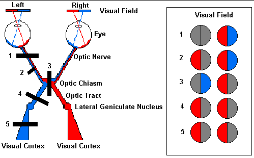
What happens if there is damage to the visual pathway? Different visual problems will occur depending on where the damage is. The black bars (labeled 1 through 5) indicate where damage may occur and the chart to the right of the pathway indicates the resulting "blind" area (gray shading) of the visual field.
Damage at site #1: this would be like losing sight in the left eye. The entire left optic nerve would be cut and there would be a total loss of vision from the left eye.
Damage at site #2: partial damage to the left optic nerve. Here, information from the nasal visual field of the left eye (temporal part of the left retina) is lost.
Damage at site #3: the optic chiasm would be damaged. In this case, the temporal (lateral) portions of the visual field would be lost. The crossing fibers are cut in this example.
Damage at site #4 and #5: damage to the optic tract (#4) or the fiber tract from the lateral geniculate to the cortex (#5) can cause identical visual loss. In this case, loss of vision of the right side.
Partial damage to these fiber tracts can cause other predictable visual problems.
The Eye | The Retina | Experiments and Activities about Vision
Hearing | Smell | Taste | Touch | Vision | Working Together
Copyright © 1996-2024, Eric H. Chudler All Rights Reserved.