Neuroscience For Kids
Brain Size
...or "My Brain is Bigger than Your Brain"
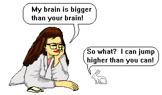 As you might imagine, larger animals have
larger brains. However, this does not mean that animals with larger brains
are smarter than animals with smaller brains. For example, a larger brain
is necessary to control larger muscles in larger animals and a larger
brain is necessary to process more sensory information from the skin in
larger animals - this has nothing to do with intelligence.
As you might imagine, larger animals have
larger brains. However, this does not mean that animals with larger brains
are smarter than animals with smaller brains. For example, a larger brain
is necessary to control larger muscles in larger animals and a larger
brain is necessary to process more sensory information from the skin in
larger animals - this has nothing to do with intelligence.
| Brain Weight (grams) | Species |
| 6,000 | Elephant |
| 1,300-1,400 | Adult Human |
| 97 | Rhesus Monkey |
| 72 | Dog |
| 30 | Cat |
| 10 | Rabbit |
| 2.2 | Owl |
| More brain weights |
 During the course of
evolution, the brain areas that show the most changes are the cerebral
hemispheres (the red areas in the drawings): the
more recently evolved animals have a larger proportion of the brain taken
up by the cerebral cortex. In the "higher" animals (especially the higher
mammals), the surface of the cerebral cortex becomes folded. This creates
grooves on the surface of the brain called sulci
(singular = sulcus). The bumps or ridges on the surface of the brain are
called gyri (singular = gyrus). The folding of the cortex
increases the cortical surface area. The cerebral cortex, made up of four lobes is involved in many complex brain functions including memory,
perceptual awareness, thinking, language and consciousness.
During the course of
evolution, the brain areas that show the most changes are the cerebral
hemispheres (the red areas in the drawings): the
more recently evolved animals have a larger proportion of the brain taken
up by the cerebral cortex. In the "higher" animals (especially the higher
mammals), the surface of the cerebral cortex becomes folded. This creates
grooves on the surface of the brain called sulci
(singular = sulcus). The bumps or ridges on the surface of the brain are
called gyri (singular = gyrus). The folding of the cortex
increases the cortical surface area. The cerebral cortex, made up of four lobes is involved in many complex brain functions including memory,
perceptual awareness, thinking, language and consciousness.
 Click on a word to hear
how it is pronounced:
Click on a word to hear
how it is pronounced:
Gyri | Gyrus | Sulcus | Sulcii
The Primary Somatosensory Cortex
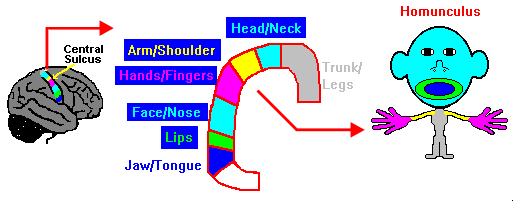
 Parts of the cerebral
cortex in the parietal lobe are involved with processing information
related to touch. One such area is the primary
somatosensory cortex which is located behind the central sulcus.
Neurons in the primary somatosensory are activated when the skin is
touched. However, the body is NOT represented in the cortex in proportion
to the amount of skin. A map of the human somatosensory cortex was drawn
by Dr. Wilder Penfield, a neurosurgeon, in the 1950s.
Parts of the cerebral
cortex in the parietal lobe are involved with processing information
related to touch. One such area is the primary
somatosensory cortex which is located behind the central sulcus.
Neurons in the primary somatosensory are activated when the skin is
touched. However, the body is NOT represented in the cortex in proportion
to the amount of skin. A map of the human somatosensory cortex was drawn
by Dr. Wilder Penfield, a neurosurgeon, in the 1950s.
After stimulating the cortex of patients undergoing brain surgery for
 epilepsy, Dr. Penfield asked the patients what they felt. By observing
the location on the brain that caused patients to feel sensations on
different parts of their bodies, Dr. Penfield was able to draw a map of
the brain. As you can see in this figure above, even though the arms and
trunk make up most of your body, they are not given much cortical tissue.
However, the face and hands take up a good portion of the primary
somatosensory cortex. This is
because the amount of primary somatosensory cortex is directly related to
the sensitivity of a body area and the density of receptors found in
different parts of the body. The areas of skin with the higher density of
receptors (like the face, hands and fingers) have more cortical tissue
devoted to them. If you were "built" in proportion to the amount of cortex
devoted to each part of your body, you would look a bit distorted: you
would have a big head and hands and a small torso and tiny legs. This
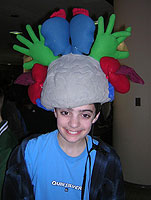
distorted body map is called a homunculus
which
means "little man."
epilepsy, Dr. Penfield asked the patients what they felt. By observing
the location on the brain that caused patients to feel sensations on
different parts of their bodies, Dr. Penfield was able to draw a map of
the brain. As you can see in this figure above, even though the arms and
trunk make up most of your body, they are not given much cortical tissue.
However, the face and hands take up a good portion of the primary
somatosensory cortex. This is
because the amount of primary somatosensory cortex is directly related to
the sensitivity of a body area and the density of receptors found in
different parts of the body. The areas of skin with the higher density of
receptors (like the face, hands and fingers) have more cortical tissue
devoted to them. If you were "built" in proportion to the amount of cortex
devoted to each part of your body, you would look a bit distorted: you
would have a big head and hands and a small torso and tiny legs. This
distorted body map is called a homunculus
which
means "little man."
 Think about how sensitive
your fingertips are compared to your leg. For a demonstration of the
sensitivity of different body areas, test your two point discrimination.
Think about how sensitive
your fingertips are compared to your leg. For a demonstration of the
sensitivity of different body areas, test your two point discrimination.
Hear it!:

- Probe the motor cortex with this science odyssey activity from PBS.
Copyright © 1996-2026, Eric H. Chudler, All Rights Reserved.