Articles and so forth
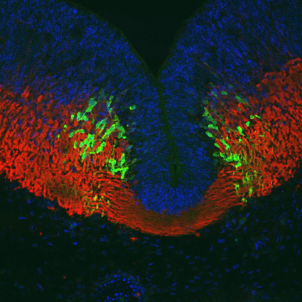
Fig. 1:Transverse section of E11.5 mouse hindbrain. The blue is nuclei stained by DAPI. Red is TuJ1-stained neurons. Green is serotonin stained with anti-serotonin antibody. The blue stained cells in the midline are ventricular zone undifferentiated cells. Just lateral to the midline are the newly differentiated serotonergic neurons of the developing raphe, which we shown are the primary drivers of spontaneous synchronized activity in the embryonic hindbrain.
This image was featured on the cover of the Journal of Physiology 550 (2003).
We study spontaneous calcium activity in embryonic mouse and chick brainstems. Spontaneous electrical activity controls many aspects of nervous system development. We have discovered that spontaneous waves of activity pass across all neurons of the hindbrain bilaterally. The description of this activity was accomplished with [Ca2+]i imaging techniques combined with retrograde labeling of identified hindbrain neuronal somata. Additional work in our laboratory has identified the midline serotonergic raphe neurons as the primary pacemakers of this activity. Furthermore, patch clamp recordings have confirmed that electrical events underlie the calcium transients and this work has also elucidated some electrical properties of the cells.
Here you can find several pages explaining aspects of the spontaneous activity.
- Page 1: About the Initial Report of Spontaneous, Synchronous Activity in Hindbrain Motor Neurons
- Page 2: Serotonergic Midline Neurons Drive Spontaneous Synchronized Activity in the Hindbrain
A complete bibliography of Dr. Bosma's publications can be found here.