Development of Spontaneous Synchronized Activity in Embryonic Mouse Hindbrain Motor Neurons
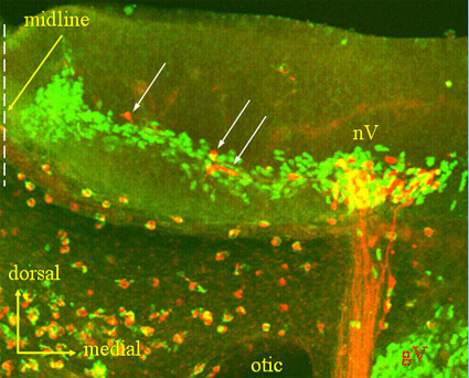
Fig. 1: Retrograde labeling of hindbrain branchiomeric motor neurons of the trigeminal nerve. Labeled axons (red) exit downward in the right portion of the picture. Their cell bodies are in the hindbrain nucleus the nerve (nV). The green color is staining for the motor neuron protein Islet-1.
From previous experiments by others, it was known that spontaneous action potentials can be recorded from emerging roots of hindbrain cranial nerves at very early embryonic stages, near the time that rhombomeric segmentation is disappearing. But the identity of the neurons in the hindbrain responsible for this activity was not known. We are able to identify hindbrain motor neurons whose axons exit in specific cranial nerves by retrograde labeling with Texas-Red-conjugated dextran injected into the target regions of the cranial nerves. Figure 1 shows this kind of retrograde label, which has identified...
After retrograde labeling, we load all neurons with the calcium indicator dye Fluo-4 and then monitor spontaneous Ca transients optically in the identified motor neurons. Figure 2 shows spontaneous Ca transients in these cells at four stages of development. Between the ninth day of development (E9.5) and E11.0, the transients become progressively shorter in duration but remain relatively unsynchronized among cells. Then, abruptly between E11.0 and E11.5, the activity becomes synchronous among cells.
Papers relevant to this topic:
- Gust J, Wright JJ, Pratt EB, Bosma MM (2003). Development of synchronized activity of cranial motor neurons in the segmented embryonic mouse hindbrain. Journal of Physiology 550(Pt 1), 123-33. Epub 2003 May 2.
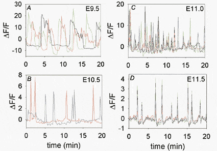
Fig 2: Records of spontaneous Ca transients from identified hindbrain branchiomeric motor neurons at four stages of development.