| |||||||
|

|
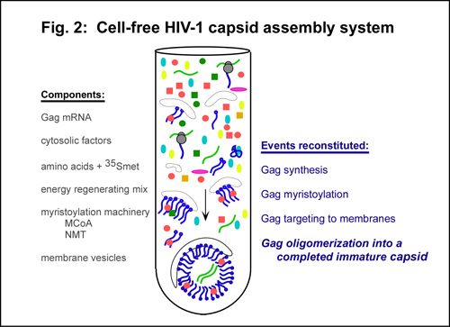
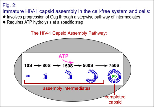
A Cell-Free System for Studying HIV Assembly: Initially, cell-free systems were used to study protein biogenesis and trafficking in normal cells and in disease. In 1991, Eckhard Wimmer first adapted cell-free systems for the study of pathogens. His group demonstrated that infectious poliovirus could be produced de novo in a cell-free system (Molla et al., 1991), proving that cell extracts could be used to reconstitute every event in virus formation, including transcription, translation, and virus assembly. However, cell-free systems are also useful for dissecting the mechanisms underlying co-translational and post-translational events. For this reason, Jaisri Lingappa, working as a postdoc at UCSF with her brother Vishu Lingappa (who was one of Blobel's first graduate students) and with Donald Ganem, developed a cell-free system to understand the mechanism by which HBV capsids are assembled in cells (Lingappa et al, 1994). This study revealed a role for cellular proteins and energy in HBV capsid formation, and directly challenged the dominant notion that capsid assembly occurs via "self assembly" in cells. Subsequently, Jaisri established a cell-free system for the assembly of immature HIV capsids, in which a HIV Gag mRNA is translated in a cellular extract containing membranes, in the presence of amino acids and energy substrates, to produce radiolabeled HIV Gag proteins. Newly-synthesized radiolabeled Gag proteins can then be tracked over time as they form capsids (Fig. 2). These experiments demonstrated that 1) formation of immature HIV capsid is faithfully reconstituted in a cell-free system; 2) Gag progresses through sequential series of assembly intermediates (identified by their sedimentation, or S, values) culminating in formation of the immature HIV capsid, and 3) energy is required post-translationally for Gag to progress through this HIV capsid assembly pathway (Lingappa et al, 1997; Fig. 3 below).
|