|
Our visual systems allow us to resolve fine detail, track a moving object,
perceive depth, and see colors. Somehow, all these components of a
visual scene merge so that we have one visual experience; for example,
when we see a cat playing with a string, we interpret the scene-paws
striking the string, details of claws and whiskers, the cat's paw in front
of or behind the string, the colors of the cat-as a unitary visual event,
even though we can attend to one or the other of these individually. How
do our brains deal with such complex visual information? First, we will
consider theories on the processing of motion, form, and color; then we
will discuss binocular vision and perceiving depth. For a brief review of
how visual information travels to the brain, see Figure 1. (For
information on the cells and connections of the visual system, see Part 1 of this unit.)
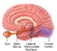 |
Figure 1. The visual pathway. Axons from ganglion cells in the
retina travel in the optic nerve to the lateral geniculate nucleus of the
thalamus. Here they synapse with other neurons, whose axons go to neurons
in the visual cortex in the occipital lobe of the brain. (Figure courtesy
of the Society for Neuroscience, copyright by Lydia Kibiuk, 1994.
http://www.sfn.org/briefings/visual_development.html) |
1. Different aspects of visual information are
processed in parallel pathways
One possibility for how we perceive a visual scene is that a defined
series of neurons and their axons handles all information-motion, shape,
and color-from a part of the visual field in a hierarchical manner. That
is, a defined set of photoreceptors, other retinal cells, lateral
geniculate cells, and cortical cells acts as a serial pathway for
information from a block of the visual scene. This information arrives at
a "master" cell or group of cells, in a visual association area of the
cortex, which combines the current information with memories of previous
experiences and make sense of it. In this model, pieces of the visual
scene are transmitted like sections of a photograph to the brain.
However, evidence does not support this "photographic transmission" or
serial pathway theory, but rather a parallel pathways model. This model
states that three primary types of information-color, shape, and
motion-are individually "pulled out of the visual scene and sent through
three parallel pathways, beginning right in the retina. In this view, one
cone receptor receives light from a small area, for example, on the face
of a moving calico cat, and signals from this cone go to several
intermediate retinal cells and from these to ganglion cells. Each of the
ganglion cells responds to only one of the attributes-color, form, or
motion-of the cat's face. The ganglion cells forward their signals to
lateral geniculate nucleus (LGN) cells in the thalamus that respond
uniquely to that one type of information (color, form, or motion), and so
on to the primary visual cortex and further cortical areas. At higher
areas, the scene is put back together again, as discussed below.
Several types of evidence, both experimental and clinical, have led to the
parallel processing theory. In one important type of experiment,
researchers recorded electrical responses from cells in specific areas of
"higher" visual cortical areas (where signals go from the primary visual
cortex). Cells in an area called V5 responded only to something moving in
part of the visual field, while cells in V4 responded to color. Second,
clinical evidence came from patients who had selectively lost the ability
to perceive one type of visual information. For example, with damage to a
particular area of the cortex, a person loses all perception of motion,
while still being able to identify objects and colors. (For a vivid
description of this syndrome, see http://www.hhmi.org/senses ; look under
"The Strange Symptoms of Blindness to Motion.") Such selective defects
are called agnosias: some of these are listed in Table 1.
| Table 1. The Visual Agnosias |
| TYPE OF AGNOSIA | SYMPTOMS |
| Object agnosia | Can't name, use, or recognize real
objects |
| Agnosia for drawings | Doesn't recognize drawn
objects |
| Prosopagnosia | Can't recognize faces |
| Color agnosia | Doesn't associate a color with an
object |
| Color anomia | Can't name colors |
| Achromatopsia | Can't distinguish hues |
| Visual spatial agnosia | Loss of stereoscopic
vision |
| Movement agnosia | Can't see objects
moving |
| Table modified from Kandel et al.,
2000 |
Putting all the evidence together, researchers now describe a system that
processes motion, the where system, and a system that handles shape or
form, the what system. The motion or where system information seems to
have its final processing station in the parietal lobe, while the shape or
what system input ends up in the inferior temporal lobe. Color
information is processed in a pathway that will be considered in Part 3 of this unit. Color information also appears to
come to its final sensory analysis in the inferior temporal lobe, but in
different cells from those for shape. From these places, messages go out
to other cortical areas-to motor systems, for example. When the brain
detects something moving, it coordinates bodily movements through the
environment or maintains eye pursuit of a moving object.
Finally, note that the parallel processing theory is still that, a
theory, and researchers disagree on how much overlap may exist in the
system.
2. Higher cortical areas reassemble the visual
puzzle
Although parallel visual processing seems to fragment what we see, at
higher levels the puzzle is reassembled. Further, the immediate visual
scene is then interpreted in light of what we know from past visual
experiences and what the wiring of our brains allows. While our visual
experiences usually make sense to us, we are generally unaware of the cues
we are using to interpret scenes until we are challenged with
unconventional pictures such as illusions. For example, when we see a
friend at some distance, we recognize the person and know that this is a
normal adult (or child) even though the image on the retina is much
smaller than that of a person standing right next to us. We are using
cues from the rest of the image, noting, for example, that the trees and
buildings also appear small, and using past knowledge to realize that
these cues mean distance, not a miniature world. When cues are not
present, we can be fooled, as shown in Figure 2. (figure 25-6, Kandel,
2000)
Figure 2.
Illusions are a window on how the brain puts together the different
aspects of visual information and integrates it with previous experiences.
Illusions presented in the Teacher Guide of this unit. illustrate higher
visual processing, but scientists do not yet know all the brain regions
and computations involved in interpreting size, shape, and motion.
3. Perceiving depth
depends on both monocular and binocular cues
Along with information on motion, shape, and color, our brains receive
input that indicates both depth, the perception that different objects are
different distances from us, and the related concept of stereopsis, the
solidity of objects. Studies show that people have two ways of judging
depth or distance: using monocular (one-eyed) information, or using
binocular (two-eyed) data. Monocular cues operate at distances of around
100 feet or greater, where the retinal images seen by both eyes are almost
identical. These cues include:
- Previous familiarity: If we know the range of sizes of people, cats,
or
trees, we can judge how far away they are.
- Occlusion: If one object partly hides another, we know that the
object
in front is closer.
- Perspective: Parallel lines such as the edges of a road, the
intersections of walls and ceilings, and railroad tracks, appear to
converge at a distance. The relative distances between objects in a scene
with parallel lines are estimated by their positions along the converging
lines.
- Motion parallax: As we move our heads or bodies, nearby objects appear
to move more quickly than distant objects; for example, telephone poles
beside the road appear to pass by much more quickly when viewed from a
moving car than do buildings or trees hundreds of feet back from the road.
- Shadows and light: Patterns of light and dark can give an impression
of depth, and bright colors tend to seem closer than dull colors.
Even though these monocular cues provide some depth vision so that the
world does not look "flat" to us when we use just one eye, viewing a scene
with two eyes-binocular vision-gives most people a more vivid sense of
depth and of stereopsis. This is important when viewing objects closer
than about 100 feet. Stereoscopic (three-dimensional) vision depends on
the fact that the eyes are separated, on average, by about six
centimeters, and thus get slightly different views of the same object.
This means that when we fixate an object (place its image on the fovea),
we can tell if another object is in front of or behind it by the
difference in location of the second object's image on the two retinas.
This difference in retinal position is called retinal disparity (Figure
3.) and is essential for stereoscopic vision. Experiments have shown that
depth perception occurs at the level of the primary visual cortex or
perhaps higher in the association cortex where individual neurons
receiving input from the two retinas fire specifically when retinal
disparity exists. Although scientists have located such neurons and know
they are important for merging or fusing the images from the two eyes,
they cannot yet fully explain how the brain accomplishes this.
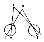 |
Figure 3. Schematic drawing looking down onto the head of someone
viewing
two objects, one indicated by the letter P and the other by the large
asterisk. The person is looking directly at the object at P, so its image
falls onto the fovea (f in the diagram). When the person notices the
other object but does not shift the direction of gaze, the image of the
other object falls on different parts of the two retinas as indicated by
the small asterisks on the retinas. This retinal disparity information is
used by the brain to interpret depth and help produce stereoscopic
vision. |
4. We first scan a scene
and then attend to individual features
Finally, what we see depends on what we pay attention to. Sometimes the
nature of the visual scene is such that one object or feature "pops out"
at us because of distinctive boundaries. If the elements of a scene are
not different enough from each other, no one part gets our attention over
another. Vision scientists describe two sequential processes that direct
our attention: the first is a rapid scanning system that tells us if a
simple property is different in one or more parts of a scene. After the
initial scan, we direct our attention to individual features of the
scene-color, shape, orientation, size-and compare each part with others to
detect subtle differences, or we compare each with a previously known
version of the scene or object. Our ability to detect a unique object in
a sea of identical objects, or to recognize any differences in what appear
initially to be two identical objects, depends on several factors, such as
the degree of differences and previous familiarity with the objects. See
Figures 3 and 4 for examples of scenes that test this ability. Students
can also devise such "minimum difference" tests. |


![[email]](./gif/menue.gif)