
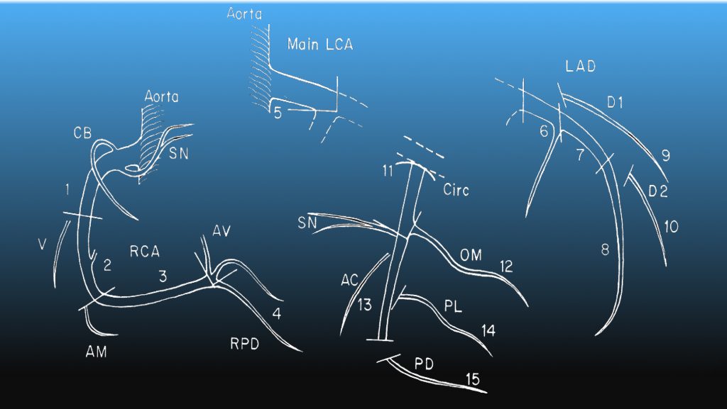
Diagram reproduced from Austen, et al.
| Segment | Description | Notes |
| 1 | Proximal Right | From ostium to ½ distance to the acute margin. Gives rise to the conus branch and Sinus node branch |
| 2 | Mid Right | Up to the acute margin |
| 3 | Distal Right | Up to the RPD origin |
| 4 | Right Posterior Descending | May be absent if left dominant |
| 5 | Left Main | Ostium to bifurcation |
| 6 | Proximal Left Anterior Descending | Up to origin of first septal perforator |
| 7 | Mid Left Anterior Descending | Up to half the distance from the first septal perforator to the cardiac apex |
| 8 | Distal Left Anterior Descending | |
| 9 | First Diagonal | May include ramus intermedius |
| 10 | Second Diagonal | |
| 11 | Proximal Circumflex | Up to and including marginal branch origin |
| 12 | Obtuse Marginal | Largest branch |
| 13 | Distal Circumflex | Distal to marginal origin |
| 14 | Posterolateral | May be small |
| 15 | Left Posterior Descending | Present in left dominant system |
References:
Austen, W. G., et al. (1975). “A reporting system on patients evaluated for coronary artery disease. Report of the Ad Hoc Committee for Grading of Coronary Artery Disease, Council on Cardiovascular Surgery, American Heart Association.” Circulation 51(4 Suppl): 5-40.
Young, P. M., et al. (2011). “Cardiac imaging: Part 2, normal, variant, and anomalous configurations of the coronary vasculature.” AJR Am J Roentgenol 197(4): 816-826.