 | Magnetic Resonance Imaging (MRI) Nets Two the 2003 Nobel Prize |
|
By Ellen Kuwana Neuroscience for Kids Staff Writer October 17, 2003 Updated: November 28, 2007 You're a doctor. Something is wrong with your patient. Don't you wish you could LOOK INSIDE your patient to see what's wrong? With magnetic resonance imaging (MRI), you can -- and the patient won't feel a thing, won't have to undergo surgery and won't be at risk for infection. Furthermore, they won't be exposed to the radiation that X-rays and CT scans require. Sound like science fiction? Until about 25 years ago, it was science fiction, but since then, the science part has caught up and overtaken the fiction part. Today, MRI is an everyday part of medicine and a valuable tool in doctors' diagnosis toolbox. When an MRI is done, a patient simply lies still in a tunnel-type device with a large magnet. The result is a detailed, three-dimensional image of the patient's insides. Doctors can tell from the MRI scan if the patient has a tumor, for example. A specialized version of this technology, functional MRI (fMRI), visualizes blood flow, so that scientists can track which parts of the brain are active during certain tasks. This type of MRI has proved to be important in studies of neurological topics such as attention deficit disorder (ADD), cancer tumor staging, pain treatment, including phantom pain, and seizure localization in epilepsy. Ongoing investigations into subjects such as decision making and language acquisition and production use fMRI techniques, as well. Now two scientists, one from the US and one from Great Britain, are being rewarded for their ground-breaking work on the development of MRI technology. They will share the 2003 Nobel Prize in Medicine, along with the $1.3 million award. Chemist Paul Lauterbur, Ph.D., joined the faculty of the University of Illinois College of Medicine as a professor of chemistry, biophysics, computational biology and bioengineering in 1985. Dr. Lauterbur passed away on March 27, 2007. Sir Peter Mansfield, Ph.D., is a professor emeritus (meaning he's officially retired, but still does some research) of the University of Nottingham's Department of Physics. |
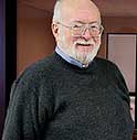 Paul Lauterbur Photo courtesy of University of Illinois Urbana-Champaign 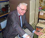 Sir Peter Mansfield Photo courtesy of University of Nottingham, School of Physics and Astronomy |
 In the 1970s, the two scientists were working independently of
each other, and made different but equally valuable contributions to the
development of MRI. Lauterbur, then a professor at the State University of
New York (Stony Brook), was using a technique called nuclear magnetic
resonance to analyze chemical samples. This technique uses magnetic fields
and radio waves of the MRI scanner to identify molecules in a chemical
sample. Interference (like static on your cellular phone) was ruining his
results. This had been a problem of this technique for more than 25 years
-- but Lauterbur came to realize that the interference could be used to
tell where atoms in the sample were located. From this information, a
two-dimensional image could be attained. In 1980, Lauterbur successfully
produced an MRI image of a patient. In the 1970s, the two scientists were working independently of
each other, and made different but equally valuable contributions to the
development of MRI. Lauterbur, then a professor at the State University of
New York (Stony Brook), was using a technique called nuclear magnetic
resonance to analyze chemical samples. This technique uses magnetic fields
and radio waves of the MRI scanner to identify molecules in a chemical
sample. Interference (like static on your cellular phone) was ruining his
results. This had been a problem of this technique for more than 25 years
-- but Lauterbur came to realize that the interference could be used to
tell where atoms in the sample were located. From this information, a
two-dimensional image could be attained. In 1980, Lauterbur successfully
produced an MRI image of a patient.Atoms are molecular building blocks. Water, for example, is made up of two hydrogen atoms and one oxygen atom (H20 is the abbreviation for water in chemistry). The human body is mostly water and fat; in fact, we're approximately two-thirds water. It's a good thing, too, or MRI would not work as well. So we're mostly water, and water is mostly made of hydrogen atoms. (The human body is 63% hydrogen atoms.) The strong magnetic fields and radio signals of the MRI align the nuclei of the hydrogen atoms. The signals that the nuclei give off vary depending on where they are in the body and on what type of tissue surround them. This exquisite sensitivity means that MRIs can detect chemical changes in tissues, thus pinpointing diseased tissue. Brain disorders generally result in a change in water content -- in fact, a change in water content of under 1% is enough to be detectable on an MRI scan. The sensitivity and precision of MRI data enable neurosurgeons to chart the path to a brain tumor, or locate areas of the brain for electrode implantation for treating Parkinson's disease.
Did You Know?
| |
|
References:
|
| GO TO: | Neuroscience In The News | Explore the Nervous System | Table of Contents |
![[email]](./gif/menue.gif) Send E-mail |
 Fill out survey |
 Get Newsletter |
 Search Pages |
 Take Notes |
 Mansfield, a
physicist, then figured out that the information could be analyzed
mathematically, thus greatly improving on the speed and efficiency of
translating the signals into an image. A rapid image enabled MRI to become
a commonplace diagnostic tool in medical centers. Mansfield said that he
had dismissed the idea years ago of winning the Nobel prize. "What you
have to do in science is to do one's best and try to survive," he said.
Sometimes, doing one's best is rewarded, as evidenced by this recognition.
MRI is used to examine almost all organs of the body, but is especially
valuable in imaging of the brain and spinal cord. It is often used in
diagnosis and treatment of
Mansfield, a
physicist, then figured out that the information could be analyzed
mathematically, thus greatly improving on the speed and efficiency of
translating the signals into an image. A rapid image enabled MRI to become
a commonplace diagnostic tool in medical centers. Mansfield said that he
had dismissed the idea years ago of winning the Nobel prize. "What you
have to do in science is to do one's best and try to survive," he said.
Sometimes, doing one's best is rewarded, as evidenced by this recognition.
MRI is used to examine almost all organs of the body, but is especially
valuable in imaging of the brain and spinal cord. It is often used in
diagnosis and treatment of