| Brain Imaging |  |
| Brain Imaging | |
 |
Brain imaging methods allow neuroscientists to see inside
the living brain. These methods help neuroscientists:
|
| Procedure | Method |
|---|---|
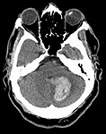
| Computed
Tomography
Scan (CT Scan)
|
CT scans use a series of X-ray beams passed through the head. The images are then developed on sensitive film. This method creates cross-sectional images of the brain and shows the structure of the brain, but not its function. |
| Positron Emission Tomography (PET)
|
A scanner detects radioactive material that is injected or inhaled to
create an image. Commonly used radioactively-labeled
material includes oxygen, fluorine, carbon and nitrogen. When this
material gets into the bloodstream, it goes to areas of the brain that use
it. So, oxygen and glucose accumulate in brain areas that are
metabolically active. When the radioactive material breaks down, it
gives off a neutron and a positron. When a positron hits an
electron, both are destroyed and two gamma rays are released. Gamma
ray detectors record the brain area where the gamma rays are emitted.
This method provides scientists with an idea of the function of
the brain. Advantages:
|
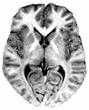
| Magnetic
Resonance Imaging (MRI)
|
MRI uses the detection of radio frequency signals produced by
displaced radio waves in a magnetic field. It provides an anatomical
view of the brain. Advantages:
|
| Functional Magnetic
Resonance Imaging (fMRI) |
Functional MRI detects changes in blood flow to particular areas of the brain. It provides both an anatomical and a functional view of the brain. |
| Angiography | Angiography involves a series of X-rays after dye is injected into the blood. This method provides an image of the blood vessels of the brain. |
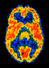
| Here are some
examples of using a combination of PET and MRI techniques:
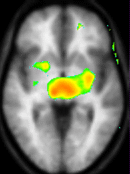 Thalamus Thalamus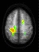 Cortex Cortex(These two PET/MRI images were provided by Dr. Robert C. Coghill at Wake Forest University School of Medicine. PET alone is also used to study different cognitive functions. |
|
For more details about MRI:
For details about functional magnetic resonance imaging:
For details about PET:
|
| BACK TO: | Exploring the Nervous System | Table of Contents |
![[email]](./gif/menue.gif) Send E-mail |
 Get Newsletter |
 Search Pages |
 Understand the relationships between specific
areas of the brain and what function they serve.
Understand the relationships between specific
areas of the brain and what function they serve.