
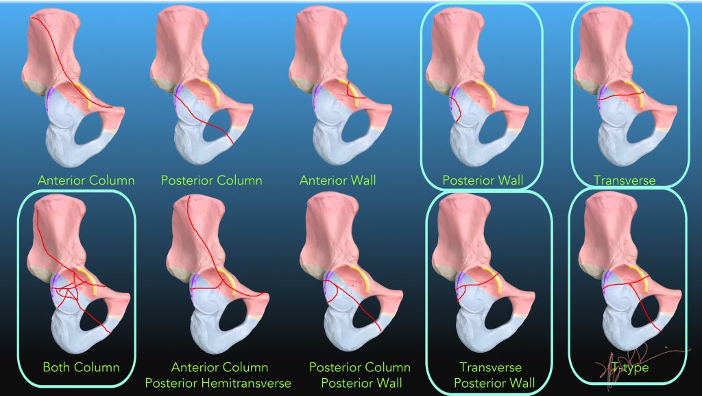
Blue frames indicate most common fracture patterns
| Classification | Description | Notes |
| Posterior Wall | Tectum intact, post wall fracture | Associated with posterior hip dislocations |
| Transverse/Posterior Wall | Sagittal tectum, post wall fractures | Associated with posterior hip dislocations |
| Transverse | Sagittal tectum fracture | |
| T-type | Sagittal tectum, obdurator ring fractures | |
| Both Column | Coronal tectum, obdurator ring fractures | Often quite complex
|
| Anterior Column/posterior Hemitransverse | Coronal with posterior sagittal tectum fractures, intact obdurator ring |
Account for most of the remainder of fractures |
| Posterior Column/Posterior Wall | Coronal tectum, obdurator ring, posterior wall fractures |
References:
Scheinfeld, M. H., et al. (2015). “Acetabular fractures: what radiologists should know and how 3D CT can aid classification.” Radiographics 35(2): 555-577.
Geijer M, El-Khoury GY. (2015). “Imaging of the acetabulum in the era of multidetector computed tomography.” Emerg Radiol 14(5):271–287.
Giannoudis PV, Grotz MR, Papakostidis C, Dinopoulos H. (2005) “Operative treatment of displaced fractures of the acetabulum: a meta-analysis. J Bone Joint Surg Br 87(1):2–9.
Judet, R., et al. (1964). “Fractures of the Acetabulum: Classification and Surgical Approaches for Open Reduction. Preliminary Report.” The Journal of bone and joint surgery. American volume 46: 1615-1646.