| Three Noble Neuroscientists |
| Three Noble Neuroscientists |
| By Ellen Kuwana Neuroscience for Kids Staff Writer October 27, 2000
And the Winners Are...Arvid Carlsson, M.D.Title: Emeritus Professor of PharmacologyUniversity of Gothenburg, Sweden Age: 77 years  Arvid Carlsson showed that dopamine was an important neurotransmitter in the
brain. Neurotransmitters are chemicals that
transmit signals from one nerve cell to another. In the 1950s, scientists
believed that dopamine was a precursor (a substance that is a building
block for another substance) for the neurotransmitter noradrenaline (also
called norepinephrine). Carlsson figured out how to measure levels of
dopamine in certain tissues in the brain, and discovered that dopamine was
concentrated in different areas than where noradrenaline was found. It
turned out that dopamine was a neurotransmitter in its own right. And
where was the dopamine? Carlsson found that dopamine was in high
concentrations in the basal ganglia, parts of the brain that are important
for movement. Arvid Carlsson showed that dopamine was an important neurotransmitter in the
brain. Neurotransmitters are chemicals that
transmit signals from one nerve cell to another. In the 1950s, scientists
believed that dopamine was a precursor (a substance that is a building
block for another substance) for the neurotransmitter noradrenaline (also
called norepinephrine). Carlsson figured out how to measure levels of
dopamine in certain tissues in the brain, and discovered that dopamine was
concentrated in different areas than where noradrenaline was found. It
turned out that dopamine was a neurotransmitter in its own right. And
where was the dopamine? Carlsson found that dopamine was in high
concentrations in the basal ganglia, parts of the brain that are important
for movement.
It is now known that dopamine levels are decreased in the brains of Parkinson's patients, especially in an area called the substantia nigra. Treating patients with L-dopa restores dopamine levels in their brain, thus lessening symptoms such as movement problems. L-dopa is still the most common treatment for Parkinson's disease. This basic research has also aided treatments for schizophrenia and depression. Paul Greengard, Ph.D.Title: Professor, Laboratory of Molecular and Cellular Neuroscience, The Rockefeller University, New York, USAAge: 74 years  Paul Greengard wanted to know how nerve cells transmit their signals. He
studied dopamine and other neurotransmitters such as noradrenaline and
serotonin to find out how they affect the nervous system. This research
was "at the molecular level," meaning that he was studying how molecules
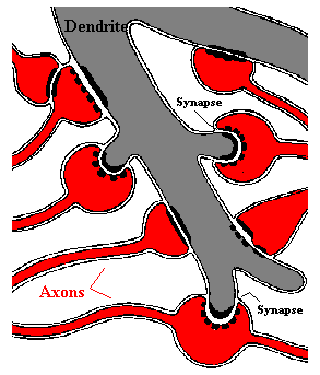
of neurotransmitter affect nerve cells, especially at the synapse. Understanding how nerve cells communicate
with each other and pass signals along has increased our understanding of
how different drugs, such as those used to treat depression and other
psychiatric disorders, work in the brain.
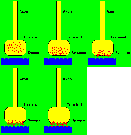
 How do nerve cells communicate? A nerve cell produces an electrical signal called an action potential. The action potential causes the release of a neurotransmitter (such as dopamine) from its end (nerve terminal). A second nerve cell responds to the neurotransmitter by producing its own electrical signal. One key event in this signaling is phosphorylation. Phosphorylation is the process by which proteins are modified by having phosphate groups attached. This changes the form, and thus the function of the protein. These proteins play a role in affecting signaling by altering the properties of the nerve cell. Phosphorylated proteins might make a nerve cell more "excitable," meaning it is more likely to send a signal. This series of events is called signal transduction, and it is how nerve cells send signals. When this signaling cascade is disrupted, for example when dopamine signaling is abnormal, disorders such as Parkinson's disease, schizophrenia, and attention deficit hyperactivity disorder can result. A deeper understanding of how signaling works under normal circumstances has helped researchers treat disorders in which signaling is abnormal. Greengard is donating his share of the prize money to a fund at The Rockefeller University for women in biomedical research. The fund is in honor of his mother, who died giving birth to him. Eric Kandel, M.D.Title: Professor, Center for Neurobiology and Behavior, Senior Investigator, Howard Hughes Medical Institute, Columbia University, New York, USAAge: 70 years  Eric Kandel, lead author of the well-known neuroscience text,
Principles of Neural Science, looked to the humble sea slug
(scientific name, Aplysia, see photograph on the left) to
investigate learning and memory at the synaptic level. Kandel was
interested in what happens to brain cells when memories are formed.
Eric Kandel, lead author of the well-known neuroscience text,
Principles of Neural Science, looked to the humble sea slug
(scientific name, Aplysia, see photograph on the left) to
investigate learning and memory at the synaptic level. Kandel was
interested in what happens to brain cells when memories are formed.The sea slug is a useful research animal because it has relatively few neurons (approximately 20,000) in its entire nervous system. Also, the neurons are always wired in the same way. However, the strength of the connections between neurons can change and be influenced by learning. Learning involves making memories, and memories bring about changes in the brain. Kandel showed that memories alter synapses in the brain. This by itself was big news, but Kandel pushed further to show that short-term memory involves changes in already existing synapses, whereas long-term memory involves creating new synapses.
Kandel wondered what was happening at the level of the synapse during learning. He discovered that there is an amplification at the synapse between the sensory nerve cell (sensing the stimulus, like your ears hear sound) and the motor nerve cell that tell muscles to perform the protective gill-withdrawal reflex. This would be like your mom calling to tell you to come home from a friend's house. Your mom's call is the stimulus, your coming home is the action--like the gill-withdrawal reflex. What is strengthened is the telephone call: maybe it is clearer, or louder. Each time she calls, you know you have to go home, and you start to learn what the phone call means. This is simple learning. The phosphorylation of proteins that Greengard studied was important for Kandel's work, too. It turns out that short-term memory involves a series of events at the synapse in which phosphorylation of proteins is key. Long-term memory has even more dramatic effects on the synapse, changing the number of proteins in the synapse, and even altering the synapse's shape! Kandel has expanded his studies to include mice. Many of the changes seen in the sea slugs are seen in mice. This suggests that this work may be applicable to humans, too. Understanding how memory formation and learning work at the synaptic level will aid in the development of drugs to treat neurological disorders such as Alzheimer's disease and dementia. References:
|
| BACK TO: | Neuroscience In The News | Table of Contents |
![[email]](./gif/menue.gif) Send E-mail | ![[survey]](./gif/menusur.gif) Fill out survey | ![[newsletter]](./gif/menunew.gif) Get Newsletter | ![[search]](./gif/menusea.gif) Search Pages | ![[notes]](./gif/menunot.gif) Take Notes |