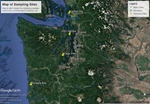
Map of sampling sites.
A map of our sampling sites.

When you think of microscopic images, what comes to mind? Cells, hairs, tiny bugs, something else? A microscope can open up a whole world of images and creatures that are too small to see with your naked eye. The microscopes we use are able to increase the perspective of something, and make it look 4, 10, 40, or even 100 times larger than it actually is. Here in the Becker Lab we mainly focus on looking at bivalve larvae, which are baby clams, oysters, and mussels. However, whilst looking for these, we come across many other fascinating specimens in their larval stages, such as: sea stars, fish eggs, copepods, snails, jellies, and barnacles. Currently, the samples we are analyzing come from the waters of Fidalgo Bay, Willapa Bay, Port Gamble, and Case Inlet (See Map of Sampling Sites for a map of these locations around Washington state).

We take photos of every single bivalve larvae we find when going through samples, but this blog post serves to show you a snapshot (pun intended) of what other marine creatures we may find when looking at a sample through a microscope. Not every sample we look at has bivalve larvae or even any other species, and we have no idea what creatures are in a sample until we open it. Therefore, every sample holds a mystery as to what we will find. Sometimes it’s nothing, and sometimes it’s something amazing.

These 6 photos show only a small portion of what we find in our samples. We find many cool creatures within the samples we look at. As aforementioned, our main focus is looking for bivalve larvae, but a fun benefit is to see what other larval creatures, be they jellies, fish, barnacles, sea stars, and more, that we can find.

Next time you’re looking through a microscope, be it at school, in a lab, at an aquarium, or anywhere else, try to take some photos of what you see, and maybe even show them to us in the comments section!
 

Bivalve larvae
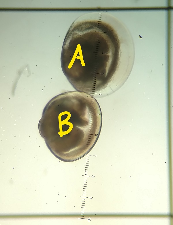
Photo 1 shows 2 commonly found bivalve larvae. Species of bivalves can look very similar in their larval stage. Thus, we have to determine subtle differences between each specimen in order to group them correctly; such as the top bivalve (A) is considered a clam due to its more rounded shape, size, and thinner shell; while the bottom bivalve (B) is a mussel because its larval shell angles to a point on one side and has a smaller bump, or umbo, than other bivalve larvae we come across.


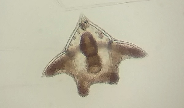
Echinoderm larva
Photo 2 shows an echinoderm larva. We’re not 100% sure what species, but after referencing images through taxonomic literature we speculate that it could be a larva of a sea star, or even a brittle star.
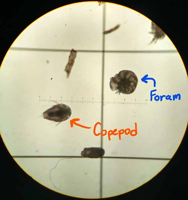
Copepod and foram in microscope.
Photo 3 shows a few other creatures that we commonly find when looking at samples, such as a foram (on the right) and an unknown species of copepod (on the left).


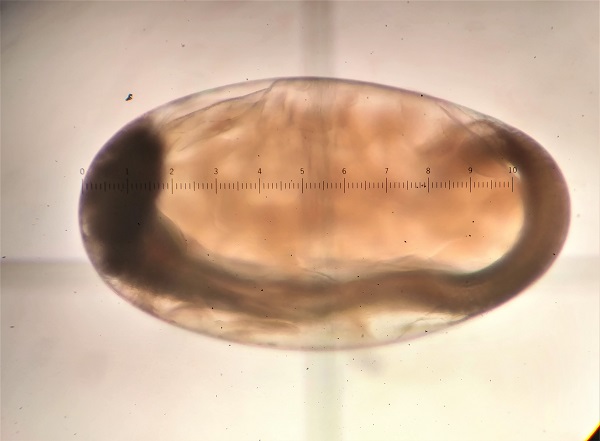
Fish egg
Photo 4 shows a fish egg. The juvenile fish is the darker part of the egg, while the lighter colored parts is the yolk sustaining it until the fish hatches.


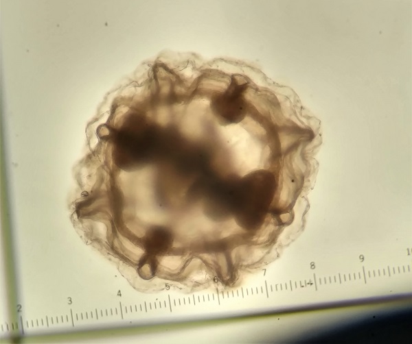
Jelly
Photo 5 shows a gelatinous-looking creature, that we think may be the top view of a larva of a jelly, aka a jellyfish. However, without being able to move the specimen around, and without seeing any tentacles, we do not have a 100% positive ID on the possible taxonomy of this creature. We commonly come across specimens that we have to ask other scientists about, which can add to the fun of not knowing what a sample may hold. If you have any ideas of what this creature may be, please let us know!


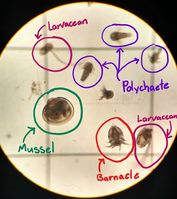
mixed plankton in scope
Photo 6 shows another view of what is commonly found in our samples alongside the bivalve larvae. In this photo, there is a mussel larva, just like the one in Photo 1, along with a barnacle Nauplius (a certain stage in the barnacles larval life), three polychaete larvae, and a couple larvaceans.

Beyond the Bivalves
Tagged on: