 |
Department of Bioengineering, Box 355061, University of Washington, Seattle, WA 98195, USA |
|
Gallery of Orphaned Graphics
OK, by now you have probably realized that I am a frustrated graphics designer masquerading as a university professor. Who says science or engineering have to be ugly? I spend a lot of time generating graphics that either are never seen or only show up as tiny versions in black and white in grant proposals. Below are links to a few of my favorites, generally using InfiniD or one of its successors. Is it science? Engineering? Art? A waste of disk space? Just wait 'till I get the movies on the site....
To see the files, click on the files listed at the left. You go direct to the .jpg files, so you have to use the "back" button to return to this menu. Note that a few of them are too big to see on a reasonable sized screen, and take a long time to download.
| 2D electrophoresis device | A design for a project that never was funded. Pity.... |
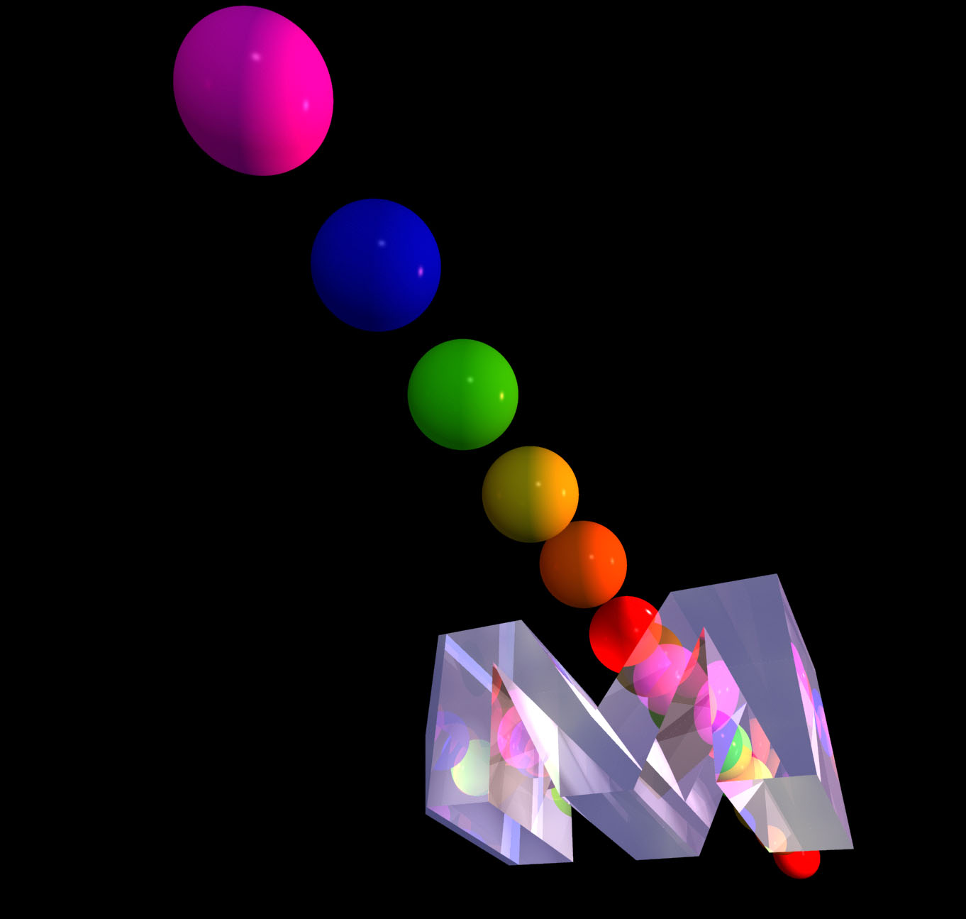
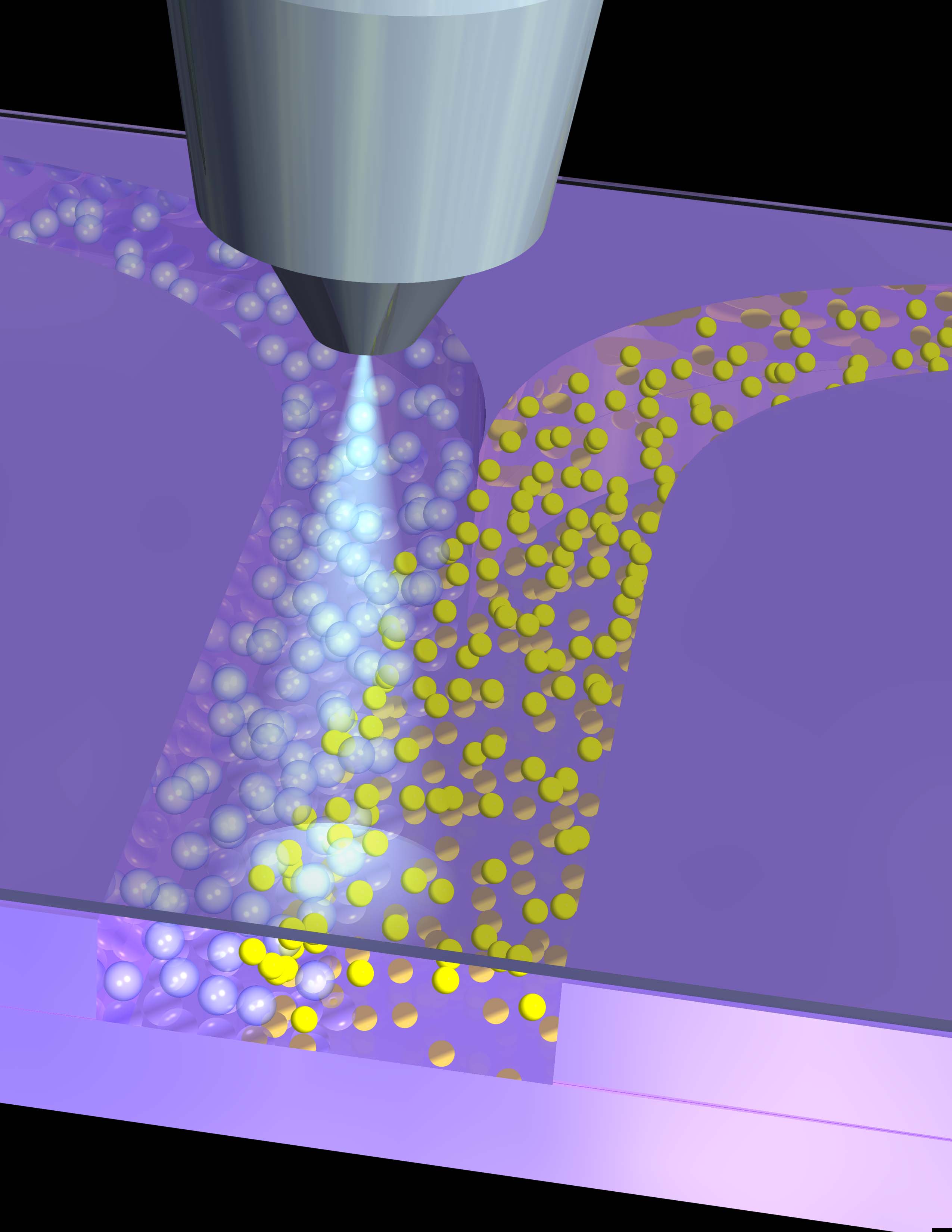
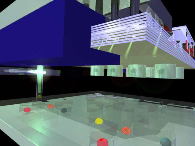
| balls and glass | Some fun with the concept of flow cytometry with V-grooves (and the first letter of Micronics) |
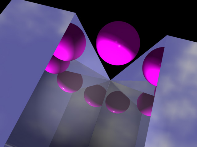
| bead in groove of M | The opening frame from a movie of the spheres marching through the Micronics M. |
| bacterium with bound antibodies | Graphics were developed to explain how one could detect whole bacteria using flow cytometry with fluorescently labeled antibodies. Voila. |
| blue molecule sandwich | A representation of a sandwich immunoassay on a bead (with a fluorescently labeled secondary antibody) |
| cascaded isoelectric focusing | A design for a device that would allow transverse isoelectric focusing with a wide range of pH values. |
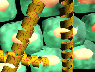
| cells and helices | An image prepared to represent the interaction of DNA-coated lipid tubules with cells for transfection |
| flow cytometer meets Julia | The Micronics laminate flow cytometer design done up pretty. |
| DIA in silicon with objective lens | This is meant to represent what the microscope sees when we image the DIA. This is a big file! |
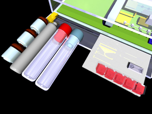
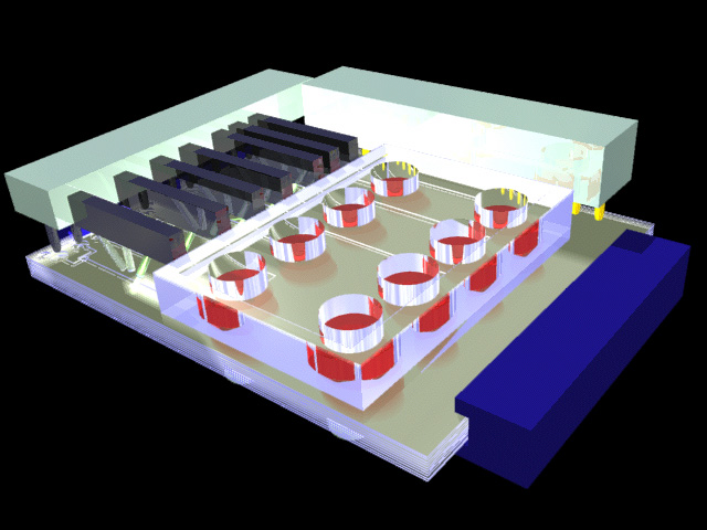
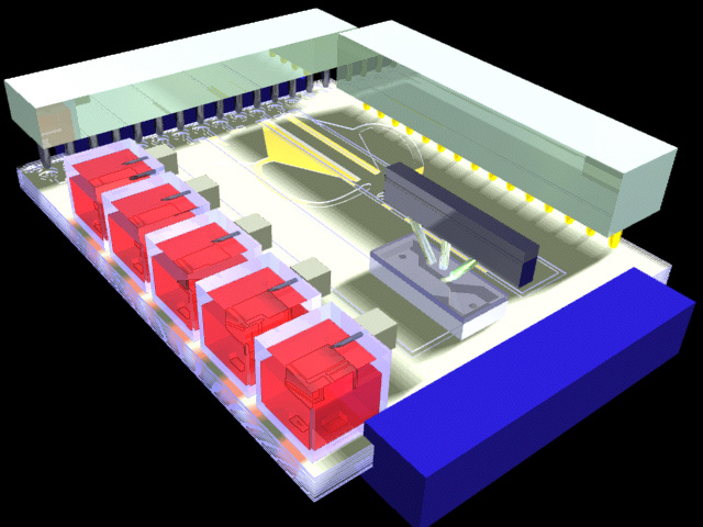
| disposables | I designed a very detailed CBW agent detection system at one point, for a project that was almost (but not) funded. It was one of the most elaborate things I have designed. |
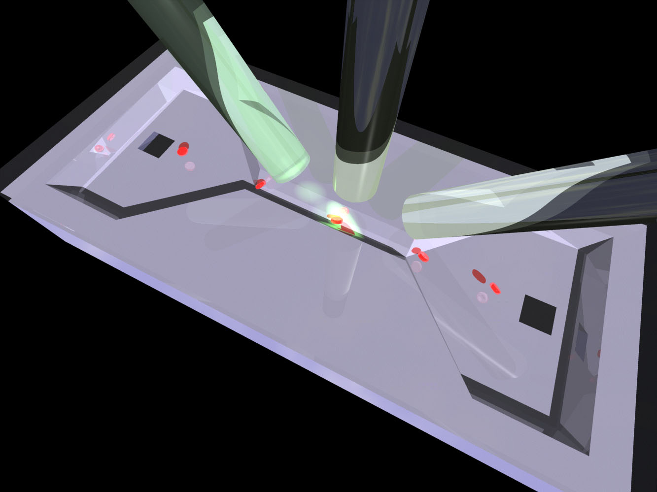
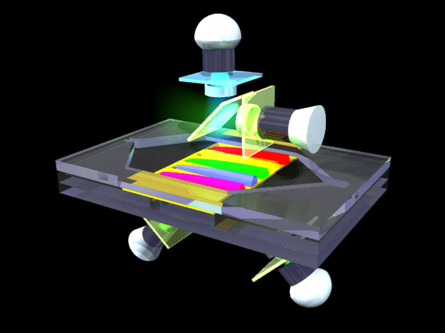
| high resolution flow cytometer | This is a V-groove flow cytometer that sat in the middle of the CBW agent detection system shown above. It exercised InfiniD's ability to hold detail at a wide range of size. Note that those are red cells in the channel. |
| IEF for cells | A design for a system for separating cells and small subcellular particles by transverse microfluidic IEF. |
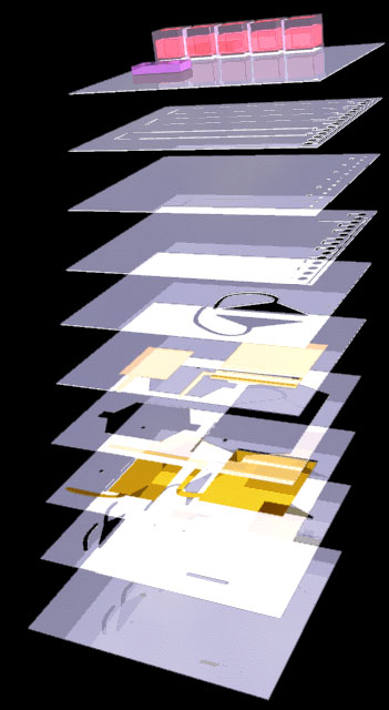
| laminate blowup | The laminate stack used in the disposable CBW agent detection system |
| another encounter of the flow cytometer with Julia and laser | Julia set under the flow cytometer again. |
| Irvine system | The proposed system for preparing nanoparticles for gene therapy. |
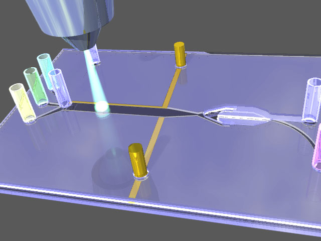
| H-filter with reservoirs | A shot of one of a set of 24 such sets of wells in a 96-well format H-filter. |
| sandwich assay | Schematic of how the assay would look? |
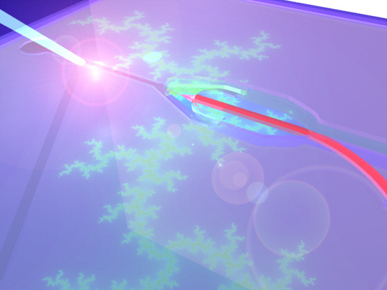
| SPR cartridge with optics | The most grandiose possible outcome of the current immunosensor system, using both SPR and fluorescence detection on opposite sides of the gold film. |
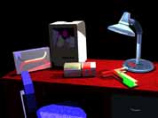
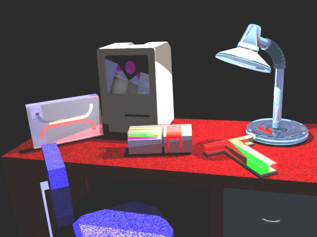
| virtual desk scene | Note that there's an H-filter, a T-sensor, a multichannel T-sensor with a blood-removing H-filter upstream, and the (original) Mac has the first image of the first sequence of a Micronics logo movie on the screen |
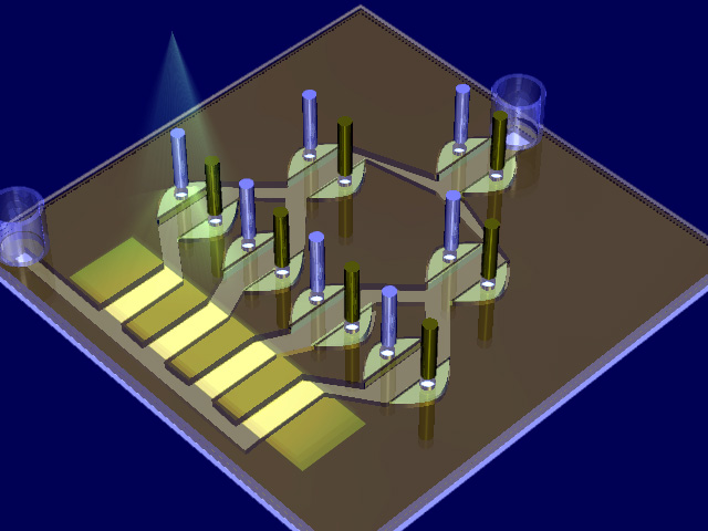
| CSI device 1 | A few years ago we had plans to design a cell handling system that could pick and place cultured cells one-at-a-time and both count and analyze them with flow cytometry. |
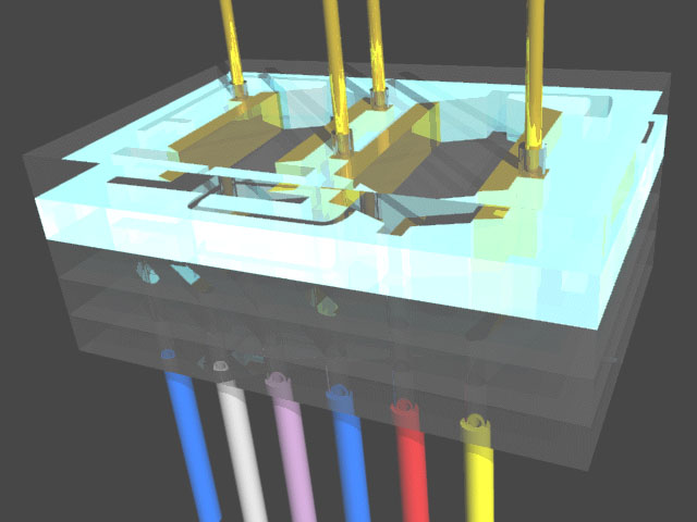
| CSI device 3 from top | More of the same--note the elctrodes a few layers down in the laminate--this design had about 12 layers, most of which you can't see here! |
| CSI devices 1&2 | And now the process of picking up those little colored eukaryotic cells with a light-monitored cannula |