Serotonergic Midline Neurons Drive Spontaneous Synchronized Activity in the Hindbrain
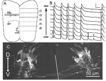
Fig. 1: A. Diagram shows schematic of open-brain hindbrain at E11.5. Former rhombomeres 2-6 are indicated on right. Trigeminal and facial motor neurons and exit points are shown on left in red. Small rectangles indicate imaging regions.
B. Intracellular Ca records from indicated regions arrayed medial (bottom) to lateral (top) demonstrating several waves of spontaneous activity that sweep across hindbrain. Note that the medial transients occur first. Open circles indicate transients that do not propagate out of the medial regions. The bottom panel shows the midline driver neurons stained with an anti-serotonin antibody. Note the axons crossing the midline, providing a basis for synchronizing spontaneous activity between the two sides of the hindbrain.
C. Immunocytochemistry for serotonin reveals a population of midline serotonin-positive neurons, with axons extending towards the marginal zone (bottom of image) and crossing the midline (indicated by line). This band of neurons is the appropriate size and in the appropriate position to play the role of midline drivers. The spontaneous activity is blocked by serotonin antagonists.
Using [Ca2+]i imaging of the entire retrogradely mapped mouse hindbrain, we have demonstrated that spontaneous activity is widespread across the entire hindbrain at embryonic day 11.5 (E11.5). This stage is the point in development where rhombomere borders disappear and synchronization is first observed between motor neurons in the hindbrain. We have demonstrated that the midline of the hindbrain drives this activity, using both high-speed propagation analysis and transaction of the hindbrain. Immunocytochemistry for serotonin shows the existence of a midline band of serotonergic neurons that corresponds anatomically to the initiating region of the midline, and spontaneous activity is blocked by application of a serotonin antagonist specific to the 5HT-2(A) receptor. We also show that this receptor is specifically localized in the marginal zone of the hindbrain, in a position appropriate for mediating the lateral spread of activity from the midline initiating zone.
Papers relevant to this topic:
- Hunt PN, McCabe AK, Bosma MM (2005). Midline serotonergic neurones contribute to widespread synchronized activity in embryonic mouse hindbrain. Journal of Physiology 566(Pt 3), 807-19. Epub 2005 Jun 2.
- O'Donovan, MJ (2005). Serotonergic neurons drive spontaneous activity in the developing mouse hindbrain ("Perspectives" on above article). Journal of Physiology 566(pt 3), 643.