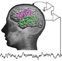
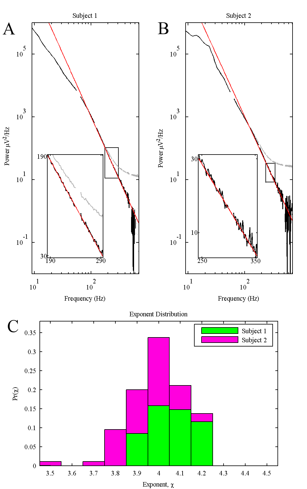
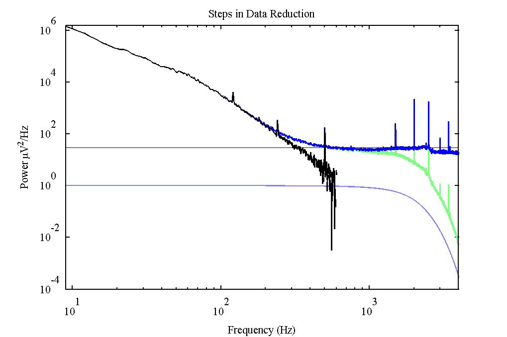
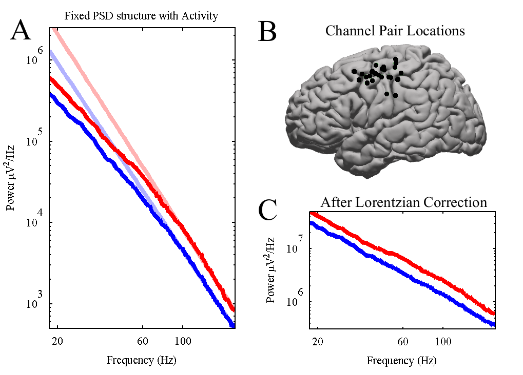
Power-law scaling in the brain surface electric potential. by Miller KJ, Sorensen LB, Ojemann JG, den Nijs M. PLoS computational biology, Vol. 5, No. 12. (18 December 2009), e1000609. doi:10.1371/journal.pcbi.1000609 Key: citeulike:6461678 Recent studies have identified broadband phenomena in the electric potentials produced by the brain. We report the finding of power-law scaling in these signals using subdural electrocorticographic recordings from the surface of human cortex. The power spectral density (PSD) of the electric potential has the power-law form P(f ) approximately Af(-chi) from 80 to 500 Hz. This scaling index, chi = 4.0+/-0.1, is conserved across subjects, area in the cortex, and local neural activity levels. The shape of the PSD does not change with increases in local cortical activity, but the amplitude, A, increases. We observe a "knee" in the spectra at f(0) approximately 75 Hz, implying the existence of a characteristic time scale tau = (2pi f(0))(-1) approximately 2 - 4ms. Below f(0), we explore two-power-law forms of the PSD, and demonstrate that there are activity-related fluctuations in the amplitude of a power-law process lying beneath the alpha/beta rhythms. Finally, we illustrate through simulation how, small-scale, simplified neuronal models could lead to these power-law observations. This suggests a new paradigm of non-oscillatory "asynchronous," scale-free, changes in cortical potentials, corresponding to changes in mean population-averaged firing rate, to complement the prevalent "synchronous" rhythm-based paradigm.
Cortical activity during motor execution, motor imagery, and imagery-based online feedback. by Miller KJ, Schalk G, Fetz EE, den Nijs M, Ojemann JG, Rao RP. Proc Natl Acad Sci U S A. 2010 Mar 2;107(9):4430-5. Epub 2010 Feb 16. Imagery of motor movement plays an important role in learning of complex motor skills, from learning to serve in tennis to perfecting a pirouette in ballet. What and where are the neural substrates that underlie motor imagery-based learning? We measured electrocorticographic cortical surface potentials in eight human subjects during overt action and kinesthetic imagery of the same movement, focusing on power in high frequency (76-100 Hz) and low frequency (8-32 Hz) ranges. We quantitatively establish that the spatial distribution of local neuronal population activity during motor imagery mimics the spatial distribution of activity during actual motor movement. By comparing responses to electrocortical stimulation with imagery-induced cortical surface activity, we demonstrate the role of primary motor areas in movement imagery. The magnitude of imagery-induced cortical activity change was 25% of that associated with actual movement. However, when subjects learned to use this imagery to control a computer cursor in a simple feedback task, the imagery-induced activity change was significantly augmented, even exceeding that of overt movement. Decoupling the Cortical Power Spectrum Reveals Real-Time Representation of Individual Finger Movements in Humans by: K. J. Miller, S. Zanos, E. E. Fetz, M. den Nijs, J. G. Ojemann J. Neurosci., Vol. 29, No. 10. (11 March 2009), pp. 3132-3137. doi:10.1523/JNEUROSCI.5506-08.2009 Key: citeulike:4169769 During active movement the electric potentials measured from the surface of the motor cortex exhibit consistent modulation, revealing two distinguishable processes in the power spectrum. At frequencies <40 Hz, narrow-band power decreases occur with movement over widely distributed cortical areas, while at higher frequencies there are spatially more focal power increases. These high-frequency changes have commonly been assumed to reflect synchronous rhythms, analogous to lower-frequency phenomena, but it has recently been proposed that they reflect a broad-band spectral change across the entire spectrum, which could be obscured by synchronous rhythms at low frequencies. In 10 human subjects performing a finger movement task, we demonstrate that a principal component type of decomposition can naively separate low-frequency narrow-band rhythms from an asynchronous, broad-spectral, change at all frequencies between 5 and 200 Hz. This broad-spectral change exhibited spatially discrete representation for individual fingers and reproduced the temporal movement trajectories of different individual fingers. home page of Marcel den Nijs, or the Physics Department URL. |