We have helped to pioneer a new medical imaging technology that combines PET and CT scanners
to produce images that show both a patient's anatomy and metabolic activity.
The combination images have proven especially effective for diagnosing cancer,
and are being incorporated into planning and monitoring treatment therapy.
PET/CT scanners will likely have other applications in areas such as cardiac imaging,
and are currently the fastest growing segment of the medical imaging market.
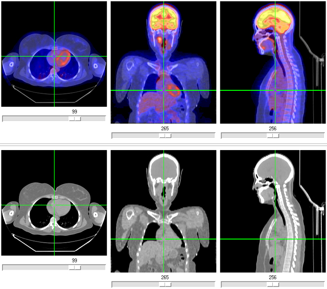
In the high-resolution x-ray computed tomography (CT) images on the bottom, denser areas, such as bone, are brighter. In the fused view in the top three panels, the positron emission tomography (PET) image is overlaid, where hotter colors represent increased metabolic activity. Increased metabolism in the heart wall and brain are clearly visible. Also visible below the brain (middle PET image) is a focus of increased activity from a thyroid cancer. The combined anatomical and metabolic information was important for the planning of the successful surgery for the patient.