Researchers
Juergen
Meyer, Eunsin
Lee, James
Eagle, Steve
Marsh, Rob
Emery, Eric Dorman,
Eric Ford
Overview
The
emerging cancer treatment approach of Microbeam
Radiation Therapy (MRT) holds promise to
revolutionize the way radiotherapy is performed.
The difference from conventional radiotherapy is
the ultra-high dose modulation on a micron scale
and consequent astonishing radiobiological
benefits. Several pre-clinical studies at a few
synchrotron facilities have shown effective
tumor response to MRT in tumor-inoculated rodent
models that are resistant to conventional
radiation therapy. Remarkably this is achieved
while preserving normal tissue functionality
following a nominally lethal high dose from the
microbeam irradiation. This “tissue-sparing
effect” makes MRT particularly suited for
treatment of pediatric brain tumors, causing
less damage to the developing central nervous
system. The extraordinary normal tissue
tolerance to highly spatially modulated high
dose MRT beams cannot be explained with our
current understanding of the radiobiological
processes and requires a paradigm shift in our
thinking. The spatial dose modulation, with
gradients of hundreds of Gray over tens of
microns, is a crucial factor in the efficacy and
safe translation of MRT to humans.
Spatially modulated proton beams theoretically
offer radiobiological potential that may provide a
dosimetric benefit over synchrotron generated
kilovoltage X-ray MRT beams, because protons have
the distinct dosimetric advantage of depositing a
large portion of their energy at depth, referred
to as the Bragg peak. By means of Monte Carlo (MC)
simulation with TOPAS, we will investigate the
feasibility of producing spatially modulated
proton beams (pMBRT) on the UW research proton
beamline that are comparable in dimensions to
synchrotron generated X-ray micro- or minibeams.
The ultimate goal of this research is to enable
radiobiology research with spatially modulated
high dose rate proton beams to be able to conduct
comparative measurements with non-modulated beams.
Guided by Monte Carlo simulations we will build a
collimator with the optimal physical
configuration.
 ' ' |
 (a)
|
 (b)
|
|
|
|
50 MeV proton beamline for
small animal research integrated into the
Small Animal Radiation Research Platform
(SARRP) at the University of Washington,
Radiation Oncology Department |
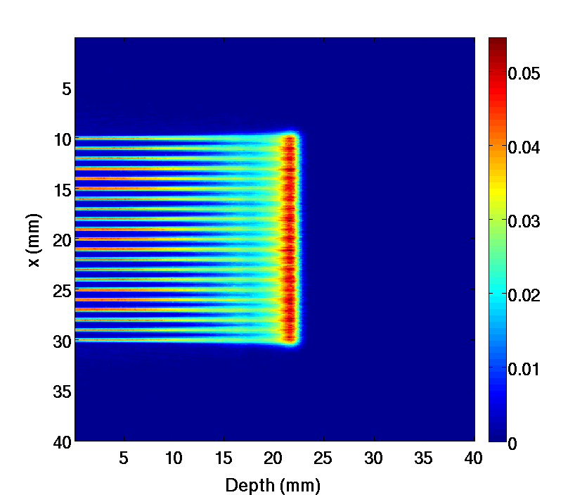
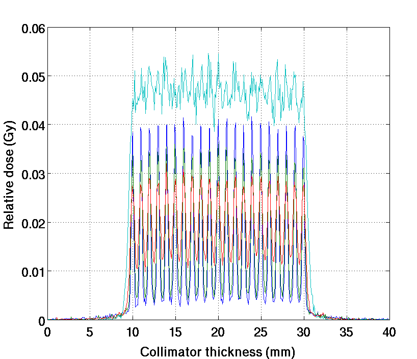
Simulated brass collimator with 0.1 mm slit, 1 mm center-to-center spacing and 5 cm thick collimator. (a) 2D depth dose showing the high modulation on the entrance side and uniform dose at depth. The color bars represent relative dose. | (b) The cross
profiles at 5, 10, 15 mm depth and through
the Bragg peak. The high modulation at
shallow depth can be clearly seen, whereas
at the depth of the tumor the dose is more
or less uniform. |
Links and Collaboration
Department of Physics and Astronomy, Medical
Physics Group, University of Canterbury,
Christchurch, NZ
References
Eunsin
Lee, Juergen Meyer, Feasibility of spatially
modulated proton beams for small animal
research, submitted to the 57th Annual Meeting
of the American Association of Physicists in
Medicine (AAPM), in Anaheim, CA, July 12-16,
2015
Back to Juergen's homepage