
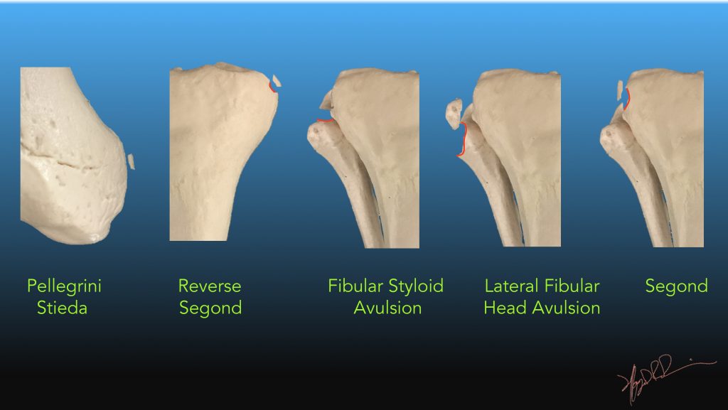
| Classification | Description | Notes |
| Pellegrini Stieda | Calcification adjacent to the medial femoral condyle | Avulsion of the origin of the medial collateral ligament |
| Reverse Segond | Small avulsed fragment from medial tibial plateau | Medial Collateral Ligament avulsion with high rates of disruption of the posterior cruciate ligament, MCL and medial meniscus |
| Fibular Styloid Avulsion | Small fragment (mm) superomedial to the styloid
|
Arcuate complex inserts on the fibular styloid. Implies posterolateral corner instability |
| Lateral Fibular Head avulsion | Larger fragment (1.5-2.5 cm) displaced 2 – 4 cn from fibular head | Conjoined tendon inserts on the lateral fibular head. Implies posterolateral corner instability |
| Segond fracture | Small avulsed fragment from the lateral tibial plateau | Lateral Capsular Ligament avulsion. High rates of Anterior Cruciate Ligament and meniscal tears |
Reference:
Gottsegen, C. J., et al. (2008). “Avulsion Fractures of the Knee: Imaging Findings and Clinical Significance.” Radiographics 28(6): 1755-1770.