| The Synapse - Up Close and Personal | |
 Developed in the 1950s, the electron
microscope
can magnify objects thousands of times. The electron microscope
passes electrons through an object and the resulting image is recorded on
film. Developed in the 1950s, the electron
microscope
can magnify objects thousands of times. The electron microscope
passes electrons through an object and the resulting image is recorded on
film. |
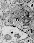
| Using the electron microscope, Dr. Pati
Irish in the Department
of Neurological Surgery at the University of Washington has taken
these pictures of synapses. The "d" represents a dendrite and the "R"
represents an axon terminal. If you look closely, you can even see some
round synaptic vesicles that contain neurotransmitters. The fuzzy black
areas represent the actual synapse between terminal and dendrite. The
larger oval objects (there are two in the dendrite of image 1 and one in
the dendrite of image 2 are "mitochondria".
| ||||||||||||
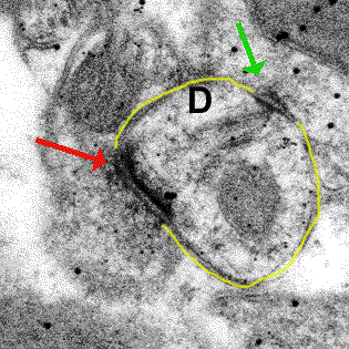 |
Photographs using the electron microscope have shown that synapses can be either asymmetrical (red arrow) or symmetrical (green arrow). In the figure on the left, notice that the red arrow is pointing to a synapse that has one dark band and one lighter band. The green arrow is pointing to a synapse that has two dark bands. Asymmetrical synapses are thought to be excitatory synapses and symmetrical synapse are thought to be inhibitory synapses. The yellow line outlines the dendrite (D). |
For more information about electron microscopy, see:

| BACK TO: | Synapses | Exploring the Nervous System |
![[email]](../gif/menue.gif) Send E-mail |
 Fill out survey |
 Get Newsletter |
 Search Pages |