|
1. Our sense of hearing
detects sound waves
Our ears alert us to events in the environment, and they detect that
special human form of communication, speech. Our hearing mechanisms
accomplish these tasks by sensing sound waves, which are changes in air
pressure, and converting these changes into electrical signals that the
brain can analyze and interpret.
2. Sensory receptors called hair cells turn air
pressure changes into neural signals
Just as we do not actually smell with the bumps on our faces called noses,
neither do we perceive sound solely with the flaps we call ears. Although
hearing begins with the ear flap or pinna, the receptor cells that change
sound energy into the electrical currency of the nervous system lie deep
inside the temporal bone of the skull. Like olfactory cells that detect
odors, auditory receptor cells (also called hair cells) are recessed from
the surface of the body. Unlike olfactory or taste receptors, however,
hair cells are not renewed when they die or are damaged. Although taste and olfactory cells interact directly with
molecules in the environment , auditory receptors are quite far removed
from the phenomena they detect. Sound waves are converted into vibrations
in a fluid in the inner ear, and these vibrations indirectly move the hair
cells, which then send electrical signals to the brain. The next few
paragraphs explore in detail the way this happens.
3. Sound activates the
external, middle, and inner ear
External Ear The folds and ridges of the pinna are not just
decorations (or for holding earrings) - they serve to channel sound
efficiently into the ear canal and to the eardrum, or tympanic membrane,
at its end. The pattern of folds captures sounds in a way that helps us
localize the origin of sound in space, especially on the vertical axis.
(Other mechanisms also help us localize sound: these will be discussed
later.) The ear canal carries sound to the eardrum, and its lining
produces ear wax to keep the eardrum and canal from drying out and to trap
dirt before it gets to the eardrum. (See Figure 1.) When sound waves
vibrate the eardrum, sound energy is transferred to the middle ear.
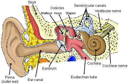
Figure 1. The main structures of the external, middle, and inner ears.
Image used with the courtesy of Dr. Beth Hartwell, University of Texas,
Houston
Middle Ear
The middle ear is a small, air-filled pocket bounded by the eardrum on one
side and the oval window of the inner ear on the other. This pocket is
connected to the common mouth and nasal cavity, or pharynx, by the
Eustachian tube (Figure 1). The Eustachian tube allows air pressure to
equalize between the outside of the eardrum (surrounding atmosphere) and
the inside of the eardrum (the middle ear). This is also the pathway that
allows infections from the mouth and nose cavities to enter the middle
ear, causing the common ear infections of childhood. The middle ear
houses the three smallest bones in the body, the malleus, incus, and
stapes (hammer, anvil, and stirrup), which form a chain of levers
connected by joints. The malleus is attached to the eardrum by ligaments,
as is the stapes to the oval window. Thus, this series of membranes and
bones forms a pathway that carries vibrations from the eardrum to the
inner ear. The stapes, the last bone in the chain, pulls or pushes on the
membranous oval window when the eardrum and the three bones are vibrated
by sound waves; the oval window is a closed membrane, but acts as the
entrance to the inner ear for sound energy.
Inner ear
What does this pulling and pushing on the oval window do in the inner
ear? A look at the structure of this area helps show how sound wave
energy is transmitted to fluid in the inner ear.
The inner ear is composed of the cochlea, from the Greek word for snail,
and the semicircular canals (Figures 1 and 2). (These latter canals are
part of the vestibular system for balance and will not be considered
here.) The cochlea is a membranous tube that is covered by a very thin
layer of bone and wound around a tiny central bone (the modiolus) into a
shape that resembles a snail; it is only about nine millimeters
across - well under one-half inch. The cochlea is filled with a special
fluid, and the pushing and pulling of the stapes on the oval window moves
the fluid in this coiled tube.
If we stretch out the cochlear tube, as in Figure 2, we see that inside
are actually three tubes, two larger and one smaller, with the small tube,
(the scala media) lying between the two larger ones. All three tubes are
filled with fluids, which vary somewhat in composition.
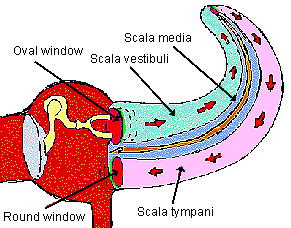
Figure 2. The middle ear and the unrolled cochlea of the inner ear. The
cochlea is a tube made of three inner tubes: the scala
vestibuli, scala media, and scala tympani. Image used with the courtesy
of http://medic.med.uth.tmc.edu/Lecture/Main/ear.htm
The fluid in the top-most tube in Figure 2 is set in motion by the
piston-like movements of the stapes on the oval window. As indicated by
the arrows, the vibrations travel into the fluid of the upper tube of the
cochlea and around the tip of the organ into the fluid of the lower
tube. The pushing or pulling of the oval window on this fluid must have a
release or dampening mechanism: this is provided by the round window, a
membrane located at the end of the lower of the large tubes in the figure.
Forming the lengthwise partition between the lower large tube and the
small tube is the basilar membrane, shown in Figure 3. On this membrane
sit the stars of the show in the auditory system, the auditory receptor
cells, or hair cells. When the basilar membrane moves,
it stimulates the hair cells, which then send signals about sounds to the
brain.
We can summarize the workings of the ear as follows:
- The pinna captures sound waves and channels them through the ear canal
to the eardrum.
- Vibrations of the eardrum pass along the three bones of the middle
ear,
with the base of the stapes then rocking the oval window in and out.
- The membranous oval window acts something like a piston in a hydraulic
system: it pushes and pulls on the enclosed fluid of the cochlea.
- The fluid vibrations move the basilar membrane, and this motion
activates auditory receptor cells (hair cells) sitting on the membrane,
which send
signals to the brain.
 |
Figure 3. Cross section of the uncoiled cochlea, showing the three
tubes and the hearing organ, the organ of Corti. Hair cells sit on the
basilar membrane and are innervated by fibers from the auditory nerve, one
of the cranial nerves. Image used with the courtesy of
http://medic.med.uth.tmc.edu/Lecture/Main/ear.htm. |
4. The basilar membrane
distributes vibrations to hair cells
The motion of the fluid in the cochlear tubes sets the basilar membrane in
motion, generating traveling waves along its length. These are somewhat
like the waves produced in a long rope that is grasped at one end and
flicked. The basilar membrane is much more complicated, though. To begin
with, it is not uniform throughout its length, but rather is relatively
wide and thin at the apex (top) of the cochlea, and narrow but thick at
the base. Because of these properties, a sound wave in the cochlear fluid
produces a peak amplitude or height of displacement of the membrane at a
certain point along its length. This point is determined by the frequency
(number of waves per unit time) of the sound that originally produced the
fluid motion. High frequencies cause a peak wave near the base (narrow
part of the membrane), and low frequencies produce their peaks toward the
apex (broad part of the membrane). Thus, the basilar membrane is
sometimes called a frequency analyzer. In addition, the hair cells on the
membrane are also tuned to particular frequencies, so that each hair cell
responds best to sound of a given frequency.
This anatomy or "geography" of the basilar membrane and hair cells
produces a tonotopic map along the membrane. This means that, as with
geographic maps, once you know some landmarks and the scale of the map,
you can calculate the point where sound of a particular frequency will
have its peak, because the system is ordered and predictable. Further,
groups of responding neurons in the brain auditory areas also contain
tonotopic maps (see below).
Figure 4. Wave traveling along the basilar membrane in the uncoiled
cochlea. For simplification, the three inner tubes of the cochlea are not
indicated. Image used with the courtesy of Dr. Fabio Mammano,
International School for Advanced
Studies, Trieste, Italy.
http://www.sissa.it/bp/Cochlea/utils/basilar.htm
5. Hair cells encode sounds
and transmit this information to neurons
So far, we have considered how sound gets to the auditory receptor
cells. But just how do these cells work? How do we tell sounds apart,
how do we recognize high and low frequency sounds, and how do we analyze
complex sounds such as human speech?
The hair cells sit on an epithelial ridge called the organ of Corti on the
basilar membrane; the ridge contains several other types of cells that
support the hair cells. The receptor cells are called hair cells not
because they sprout hairs, but because their apical or top ends are
covered with cilia, which under the microscope look a bit like hairs. Over
the top of the cilia lies a gelatinous membrane, sandwiching the hair
cells between itself and the basilar membrane. The complex, relative
movements of these two membranes activate the cilia of hair cells, causing
the cells to undergo a change in the electrical potential across their
cell membranes. When specific changes occur in this electrical state,
neurotransmitter molecules are released from the bottom or basal parts of
the hair cells. Thus, the cilia are essential in transducing, or changing,
the mechanical energy of the basilar membrane into electrical changes in
the hair cells. As mentioned above, hair cells are tuned to the
particular frequencies that activate the portion of the basilar membrane
where they reside.
Hair cells are modified epithelial cells and do not have dendrites and
axons as neurons do, but they communicate, as many neurons do, by
releasing neurotransmitter. They release the neurotransmitter at junctions
or synapses that they form on branches from neurons whose cell bodies are
in a ganglion (group of neurons) just outside the cochlea. The axons from
the ganglion neurons form the auditory nerve, which carries signals into
the first stop in the brain, the cochlear nucleus.
6. Sound information from each ear is
distributed
to both sides of the brain
Once information from one ear goes to the cochlear nucleus on that side of
the head, the neurons in this nucleus send information to identical higher
centers on both sides of the brain. As shown in Figure 5, some of the
processing stations are the superior olivary nucleus, inferior colliculus
(in the midbrain), medial geniculate nucleus (in the thalamus), and the
auditory cortex. To avoid confusion, Figure 5 shows only the pathway on
one side from the brainstem to the midbrain. From the auditory cortex,
messages go to other areas of the cerebral cortex for interpretation of
the meaning of sounds.
 |
Figure 5. Auditory pathways in the brain. Signals from
neurons that get information directly from hair cells travel in the
auditory nerve to the brainstem. Here the signals activate more neurons,
which send the auditory messages on to the thalamus, then to the auditory
cortex in the temporal lobe of the cerebrum. Image used with the courtesy
of Dr. Remy Pujol, University of Montpellier, France
In the cochlear nucleus, the first brain relay station for sound, signals
encoding sounds are not just passed on, but rather are "dissected" and
sorted first. This means that different features of a sound, such as
frequency, intensity, or onset and offset (beginning and ending of a
sound) are carried to higher brain centers separately. This sorting out of
the features of stimuli and sending messages forward in parallel nerve
pathways is a common and important attribute of brain sensory
systems. One of the big tasks of researchers is to find out how areas
in the cerebral cortex use input from these parallel pathways to interpret
the original sensations - in this case, the original sounds.
|
7. We need two ears to locate a sound
source
When a sound occurs at the extreme left of a subject, the arrival of the
sound at the left ear is about 600 to 700 microseconds (millionths of a
second) earlier than at the right ear. Further, the head acts as a sound
barrier, so the sound is a little louder in the left ear. How does the
timing and intensity of sound in the two ears tell the brain where the
sound source is?
This processing is carried out in the superior olivary nucleus (SON) of
the brainstem. Axons coming into the SON from the cochlear nuclei form
synapses successively across a linear series of SON neurons, as shown in
Figure 6. Each neuron here gets messages from cochlear neurons in both
ears, and in order to fire a signal to higher brain centers, each must
receive simultaneous messages from the two cochlear nuclei. Because a
sound from, for example, the extreme left side of a person arrives later
in the right ear than the left, hair cells and neurons from the right ear
send their signals slightly later than those from the left ear. Each SON
neuron is activated only by simultaneous input from the two ears, so that
when, for example, signals from axon d1 from the right and d2 from the
left ear coincide on SON neuron d, it fires. Through experience, we learn
that when neuron d fires, the sound is, for example, at 50o to
the left of
straight ahead. If neuron b fires, this could mean that the sound
originates at 20o to the left.
Neurons in another part of the SON employ intensity cues rather than
arrival-time cues. Again, the neurons need simultaneous input from the
two ears to fire, but in addition, they respond best when the sound
intensity on one side of the head exceeds that on the other by a certain
amount.
Note that it is hard to differentiate sounds coming from directly in front
of you from those originating directly behind you. Both sounds are
equal distance from the two ears, so there is no difference in timing and
intensity, information that our brains need to localize sound in the
horizontal dimension. In the experiments in the Student and Teacher
Guides accompanying this unit, students see how this mechanism works.
 |
Figure 6. Schematic of how sound is processed in the superior olivary
nucleus (SON). A sound arriving earlier at the left ear elicits signals
more quickly in the SON than those from the right ear. At some point, as
the signals from the two ears travel across the linear array of neurons in
the SON, they converge on one neuron and activate it (neuron "d" in this
illustration). This processing is carried out simultaneously in the left
and right SONs, but only one is shown here. |
8. Auditory brain centers contain tonotopic
maps
The frequency mapping of sounds along the basilar membrane is carried
throughout the auditory brain centers. This means that neurons in the
cochlear nucleus that respond to progressively higher frequencies are
found in an orderly progression along an axis of the nucleus. The
superior olivary nucleus, midbrain centers, and auditory cortex also
contain tonotopic maps.
Maps for locations of sound origins are also established in certain brain
centers. These are spatial maps, similar to such maps for the touch
system, and are found in groups of neurons separate from those forming
tonotopic maps.
9. Human brains decode the complex sounds of
speech
A composite sound such as a vowel sound in human speech usually has three
dominant frequency components. The movement of the eardrum and ear bones
receiving such a sound is very complex, but when the sound reaches the
basilar membrane, the frequency components are sorted out. Each of the
frequency components sets off a separate travelling wave and each wave
produces its peak at the position on the membrane that responds best to
that frequency. Next, the hair cells on the membrane at each peak send
signals indicating to higher centers that a certain frequency of sound has
been detected.
What happens when signals from language sounds are sent to higher
centers? So far, this has not been an easy question to answer. When
researchers try to find which cortical centers and cells are involved in
neurological functions, they often inject tracer materials into animal
brains, perform experiments, and then dissect the brain to find out what
areas were affected. Language processing has been difficult to study
because it is a uniquely human trait and such experiments cannot be done
on people. Studies of sonar and echolocation in bats have provided many
insights into the processing of complex sounds.
Another way scientists find out about brain areas used in language is to
study people with language problems. They then either study those
patients' brains after natural death, or before death with techniques such
as magnetic resonance imaging (MRI), which allows them to see the damaged
areas and hypothesize about the functions of those areas.
Researchers are now also using positron emission tomography (PET) to study
brain function during language processing in normal individuals. PET is a
non-invasive procedure that shows local changes in blood flow and
metabolism that occur when the brain is working-in this case, working to
interpret spoken language. Through studies such as these, scientists have
discovered many brain subdivisions for processing different aspects of
language. The area for comprehending spoken language, for example,
contains separate areas for decoding the meaning of words and for
understanding the relationship of words in a sentence. These studies are
opening new windows on how we decode language.
10. Hearing abilities vary among
animals
The ability to hear is not found as widely in the animal kingdom as some
other senses (e.g., touch, taste and smell); it is restricted mainly to
vertebrates and insects. Within these, mammals and birds have the most
highly developed sense. Animals are able to hear over quite a range of
frequencies: recall that frequency means the number sound waves per unit
of time, usually termed cycles per second or hertz (Hz). Here are some
examples of the frequency ranges that can be heard by different
animals:
        	Humans:  	20- 20,000 Hz
        	Whales:    	20 - 100,000 Hz
        	Bats:         	1500 - 100,000 Hz
        	Frogs:       	600 - 3000 Hz
        	Fish:          	20 - 3000 Hz
        	Crickets:   	500 - 5000 Hz
11. Problems in different
parts of the auditory system can cause deafness
Conductive and sensorineural hearing loss
About 28 million people in the United States have hearing loss significant
enough to interfere with understanding conversations, and about one third
of people over the age of 75 have difficulty hearing. Doctors generally
divide hearing loss into conductive and sensorineural loss. The first
occurs when something happens to the ability to transfer sound waves from
the outer to the inner ear. The second results when damage occurs to the
inner ear (frequently to the hair cells) or the auditory nerve leading to
the brain. These two types of loss are both types of peripheral deafness,
meaning they are caused by problems outside the central nervous system
(brain). Central deafness is rare, because as pointed out earlier, once
sound information reaches the brainstem, it is sent to many stations on
both sides of the brain. In order for significant hearing loss to occur
from damage to these redundant areas, the brain injury would be so
extensive that many other functions would also be disrupted.
What causes deafness?
Tumors, objects in the ear canal, or repeated ear infections that damage
the eardrum can cause conductive hearing losses. Another cause of
conductive loss is otosclerosis, a genetic condition that causes the
middle ear bones to degenerate. Infectious diseases such as German
measles, mumps, meningitis, and syphilis can cause sensorineural
deafness. High blood pressure, multiple sclerosis, and diabetes can also
lead to this type of hearing problem, but most sensorineural
deafness-probably half of that found in children-is genetic, with large
extended families affected. Exposing your ears to loud noises can also
lead to sensorineural hearing loss by destroying the hair cells of the
inner ear. Further, high doses of some medicines can damage hair cells
and nerves, as can trauma from accidents.
Treating hearing loss
If the underlying cause of conductive hearing loss is treated, hearing can
often be restored. For example, treating allergies and infections can
reduce swelling and fluid in the ear, and replacing middle ear bones with
tiny metal prostheses can greatly improve hearing in cases of
otosclerosis.
But antibiotics and surgery cannot treat sensorineural loss: if loss is
not complete, a hearing aid may help-these contain a tiny microphone and
amplifier to increase volume, and now many are small enough to fit into
the ear canal. For profound (complete) loss in both ears, some people
have turned to cochlear implants that can help people hear.
What can you do to protect your hearing?
At this time, help for genetic forms of deafness are limited (i.e.,
cochlear implants; metal replacement for ear bones). For other causes,
people should consult a doctor when an ear infection occurs or when
symptoms such as pain in the ear, constant ringing, dizziness, discharge,
or bleeding occur. Another critical behavior for preventing hearing loss
is to avoid exposing ears to loud noises, either by avoiding noisy
situations or by using earplugs or ear covers. High noise levels damage or
kill hair cells. Decibel levels that damage hearing can be found at
http://faculty.washington.edu/chudler/bigear.html.
|


 [Back to Top]
![[email]](./gif/menue.gif)