Specifications for Flow Through Wood Fiber Kappa Number Analyzer
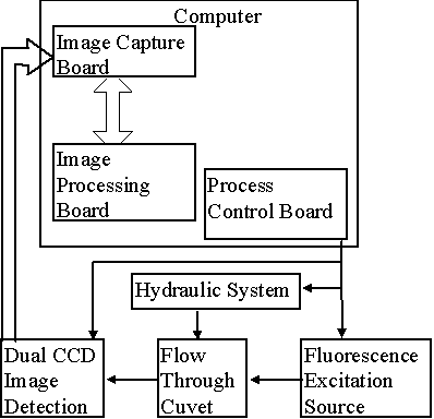
A block diagram of the proposed instrument is given in Figure 1. The wood fibers are suspended in an aqueous solution containing a fluorescent dye, acridine orange, which binds strongly to the fibers (1). Acridine orange, excited in the blue region of the spectrum at approximately 400-485 nm, fluoresces both in the green (520 - 580 nm) and red (590 - 650 nm) regions of the optical spectrum. When the suspension of fibers is examined under the fluorescence microscope at low magnification, the color of the fluorescence varies from green, indicating a low kappa number to a red, indicating a high kappa number (Figure 2).
In order to quantitatively estimate the kappa number from a fluorescence measurement, the fibers are caused to flow past a measuring station where the ratio of their integrated red and green fluorescence is determined. The flow through optical cell is similar to that described by Olson et al. (2). A close-up of the flow cell is given in Figure 3. The central flow stream containing the fibers is ensheathed in two outer streams which narrows the central stream and centers it as a sheet in the middle of the flow cell. The Reynolds number of the system is low enough so that the flow is entirely laminar and streamlines do not mix, except (slowly) by diffusion. The advantages of a sheath flow system are well known from cell cytometry (3): (a) the fibers are all oriented and in focus, and (b) the system tolerates large particles without blocking.
The optical measuring system is given in Figure 4. It is essentially a low magnification fluorescence epi illumination microscope capable of measuring two fluorescence images of the fiber simultaneously, one in the red and one in the green. A critical element is the objective. It should have a large numerical aperture and wide, flat field of view. Nikon makes a lens in the CF160 series that would seem ideal for this application. As light source, we are considering a pulsed system to avoid problems with image blurring. The new generation of Blue LEDs may prove optimal for this application; alternatively, we may resort to pulsed Xe arc lamps.
CCD cameras will be used as the imaging devices. These will operate at 30 Hz frame rate in non-interlaced mode. The cameras should be equipped with a shuttering capability to minimize contributions to the image from stray light in the system. Image acquisition will require two independent frame grabbers, one for each CCD. It is desired that the image processing take place in real time. Image analysis will consist of the following steps: (a) scene segmentation in which the fibers will be located and isolated from the background. (b) selection of fiber images that are fully in the field, (c) integration of the fluorescence intensity of each fiber, (d) matching of fibers in the separate red and green images, (e) computation of the ratio of integrated intensities for each fiber, and (f) calculation of kappa number of each fiber and formation of a histogram of fiber kappa numbers.
References
1. Liu, Yue, The Development of a Single Fiber Kappa Analyzer, Ph. D. Thesis, University of Washington, Seattle WA, 1998.
2. Olson, J. A., Robertson, A. G., Finnigan, T. D. and Turner, R. R. H., An Analyzer for Fibre Shape and Length. Journal of Pulp and Paper Science, 21 (11) J367-373.
3. Malamed, M. R., Lindmo, T. and Mendelsohn, M. L., Eds., ‘Flow Cytometry and Sorting’, 2nd Ed., Wiley, New York, 1991.
Figure 1. Block Diagram of the Flow Through Wood Fiber Kappa Number Analyzer.
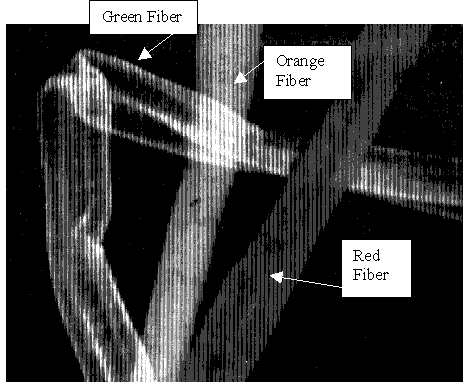
Figure 2. Acridine Orange Stained Wood Fibers of Varying Kappa Number. Low kappa number fibers are stained green, intermediate number fibers, orange and high kappa number fibers, red.
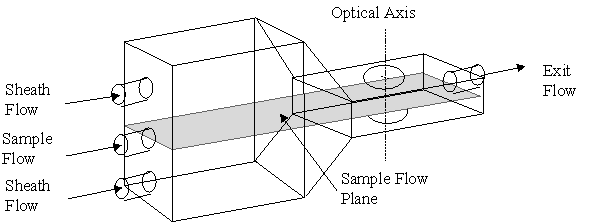
Figure 3. Flow Through Optical Cell
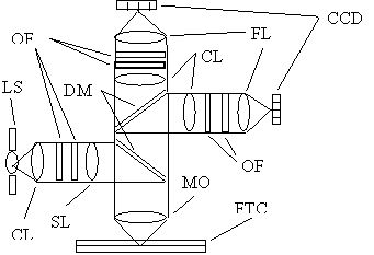
Figure 4. Optical Schematic of Flow Through Wood Fiber Kappa Number Analyzer. CCD: charge coupled device camera, CL: collecting lens, DM: dichroic mirror, FL: focusing lens, FTC: flow through cuvet, LS: light source, OF: optical filter, SL: beam shaping lens.