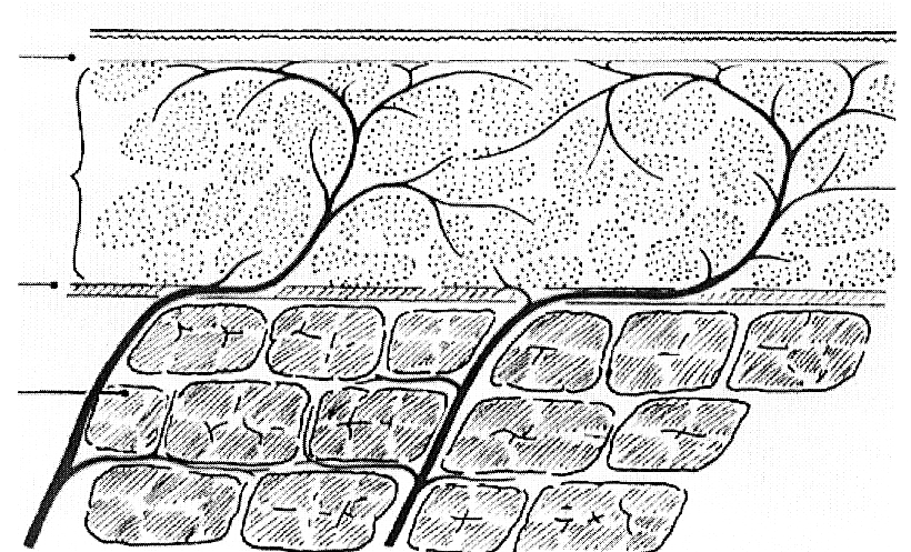
Figure 6.
From Cormack and Lamberty {2081}.
Schematic of arteries supplying skin in cross section. The
lowest layer (cross hatched cells) is skeletal muscle. The skin
layer is represented by the thin unshaded layer at the top, bounded by two
horizontal lines (for scaling take this as 1 mm thick). Between
them is a rippled line, just below the top line; this represents
the boundary between the upper epidermis and the dermis, the two
layers of skin. Between the skin and muscle layers is the
subcutaneous layer, cells stippled, on a basement membrane layer
(cross hatching between continuous horizontal lines). The
perforating arteries arise through the muscle layer without
diminution in diameter. See Cormack and Lamberty's book for
detailed renderings of varieties of perforators, such as those
that pass through fascial planes rather than muscle, and others
that travel in the subcutaneous layer.