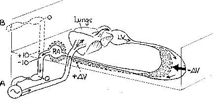
Figure 2. Schematic of physical arrangement for setting central venous pressure.
Venous return, intercepted via a cannula in the right atrium, was pumped into the pulmonary artery. Between the right atrium and the pump was a section of flexible tubing and a "Starling resistor". The latter is formed with a length of flaccid tubing so that it collapses if intramural pressure becomes even slightly subatmospheric. It serves as a visual indicator and controller of pressure, a convenient means of verifying that pressure is zero at the inflow end.
In solid lines, the resistor and connecting tubing are shown as located 10 cm below the reference level of the heart. With zero pressure at the entrance to the Starling resistor, this means that pressure at the right atrium is -10 cm. In dashed lines, the assembly is shown elevated 10 cm above the heart. After appropriate adjustments of pump rate, pressure at the entrance to the Starling resistor is again zero. Five centimeters below, pressure in the right atrium is therefore at + 10 cm.
In the schematic representation of the peripheral vasculature, the solid lines represent the volume distribution in the venous vasculature when pressure in the right atrium was -10 cm. With this low pressure in the large collecting veins, represented as the segment nearest the heart, distending pressure is low and volume within the segment is correspondingly low. Pressures are relatively high in the peripheral veins, shown bulged out in the representation of volume in the peripheral vasculature.
After elevation of the Starling resistor to produce positive pressure in the right atrium, the distribution of flow is altered in the direction suggested by the dashed lines. The volume in the segment near the heart is relatively large, increased by
D V, due to the larger distending pressures. At the low level of cardiac output associated with the condition of high central venous pressure, pressures are relatively low in the peripheral venous vasculature; the dashed line suggests the corresponding reduction of volume by DV . Changes of volume in the relatively incompliant arterial vasculature are ignored.